Case Report

Gangrenous Sigmoid Volvulus Complicating Pregnancy
*Mumtaz Din Wani,**Shabir Ahmad Mir,
- *Postgraduate Department of Surgery, Government Medical College, Srinagar, India
This is an Open Access article distributed under the terms of the Creative Commons Attribution License ((http://creativecommons.org/licenses/by/3.0)which permits unrestricted use, distribution, and reproduction in any medium, provided the original work is properly cited
Abstract
Sigmoid volvulus is a very rare cause of intestinal obstruction in pregnancy. The gangrene results due to delay caused by conservative trial for sake of pregnancy, avoidance of radiology based investigations, and rarity of the condition and the masking of clinical picture by pregnancy. Our patient was a 28 year old female with 26 weeks of pregnancy. She presented with 3 day history of pain abdomen, obstipation with 2 episodes of vomiting and distension abdomen. She also had abdominal tenderness. Her general condition was ill. She was explored after resuscitation and the exploration revealed gangrenous sigmoid volvulus with 2 ½ twists. We performed the colostomy with the closure of the rectal stump (Hartmann’s).
Case Report
A 28 year old female, gravid 4, para 3, presented at 26 weeks of gestation with 3 day history of abdominal pain, obstipation, 2 episodes of vomiting, abdominal distention and tenderness. Her pregnancy otherwise had been uneventful. She had no previous significant medical or surgical problems. On examination she was febrile with distention and tenderness abdomen. Her fundal height corresponded to 26 weeks of gestation, her preoperative vitals were BP 138/82mmHg and pulse 96bpm.
Preoperatively patient was kept nil per orally with Ryle’s tube suction and on intravenous fluids, intravenous antibiotics and proctocolysis enema but there was no improvement in symptoms. Preoperatively her laboratory parameters were: white blood cells count 9480 cell/cumm, hemoglobin12.3gram/dL, platelet count 1,35,000/cumm, blood urea 28mg/dL, serum creatinine 0.9mg/dL, blood sugar (F) 118mg/dl, sodium 141 mmol/L, potassium 3.9mmol/L. Ultrasonography of abdomen showed distension of intestinal loops with significant interloop fluid suggestive of subacute intestinal obstruction. On digital rectal examination, rectum was empty, tone was normal with no abnormality detected. The surgical team decided to explore the patient after consulting the obstetric team. Patient was explored under the cover of isoxsuprine infusion (tocolytic) and injection progesterone 200mg IM after obtaining an informed high risk consent with respect to pregnancy and low saturation (SPO2 92% on room air). Isoxsuprine infusion was continued for 24 hours preoperatively.
Intraoperative findings noted were complicated (gangrenous) sigmoid volvulus (Figure 1), about 500ml of foul smelling free fluid in peritoneal cavity and subphrenic spaces, note was also made of gravid bulky uterus. Resection of the gangrenous colon was done along with closure of rectal stump (Hartmann’s procedure) (Figure 2). Warm normal saline washes were given. Drain was kept in peritoneal cavity which was removed after about one week of surgery. Drain was kept in view of possibility of bleeding.
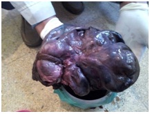
Figure 1: Resected specimen
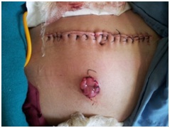
Figure 2: Postoperative picture of the patient
Postoperative parameters were pulse 100bpm, BP 110/70mmHg, antiseptic dressing was applied on alternate days. Postoperative ultrasonography for fetal well being showed transverse lie, fetal heart sound present, adequate liquor, gestational age 26 weeks and placenta-anterior / fundal. On first postoperative day, oral isoxsuprine and progesterone was started by obstetrician.
DISCUSSION
Sigmoid volvulus has a variable geographic distribution, being extremely common in developing countries where it affects the young patient, with a lower incidence in Western countries where it predominantly affects the elderly. Chronic constipation is blamed for the Western type of sigmoid volvulus, while a high amount of fiber in the diet has been deemed a major factor in the development of sigmoid volvulus in the African population [1].The sigmoid volvulus is due to a long and wide mesosigmoid that rotates on a constant mesosigmoid root width [2].
The predisposing causes of sigmoid volvulus include narrow attachment of pelvic mesocolon, long pelvic mesocolon, band of adhesions (peridiverticulitis), chronic constipation and high residue diet [3].
SV usually occurs in institutionalized, debilitated; chronically patients who have long redundant sigmoid colon [4]. The incidence of intestinal obstruction in pregnancy ranges from 1 in 1500 to 1 in 66431 deliveries [5]. Intestinal obstruction in pregnancy can be caused by many factors including congenital or postoperative adhesions, volvulus, intussusceptions, hernia and appendicitis [6]. Sigmoid volvulus is an important cause of bowel obstruction complicating pregnancy, accounting for up to 44 per cent of cases [7]. Pregnancy itself is considered to be the precipitating factor for sigmoid volvulus. The occurrence of sigmoid volvulus in pregnancy is thought to be due to displacement, compression and partial obstruction of a redundant or abnormally elongated sigmoid colon by the gravid uterus [8]. This could probably explain the increased incidence of sigmoid volvulus in the third trimester of pregnancy [6]. The average time from the onset of obstructive symptoms until presentation has been reported to be 48 hours [6]. This is largely because pregnancy itself masks the clinical picture since abdominal pain, nausea, and leukocytosis can occur in an otherwise normal course of pregnancy [9]. The maternal and fetal outcome in sigmoid volvulus has been directly related to the degree of bowel ischemia and subsequent systemic sepsis. This observation highlights the fact that high index of clinical suspicion is vital in cases of intestinal obstruction in pregnant patients. This facts needs to be emphasized amongst the general practitioners and community obstetricians primarily responsible for taking care of these patients.
Another important area of concern is the reluctance in the utilization of modern radiological diagnostic tools in pregnant patients. There have always been concerns about the radiation exposure of the fetus during pregnancy. Significant radiation exposure may lead to chromosomal mutations, neurologic abnormalities, mental retardation, and increased risk of childhood leukemia. Cumulative radiation dosage is the primary risk factor for adverse fetal effects, but fetal age at exposure is also important [10 11 12]. Exposure during the first week of gestation results in highest rates of fetal mortality. The next most sensitive time period is between 10 and 17 weeks of gestation, when central nervous system teratogenesis becomes an important consideration. After this period, the concern shifts from teratogenesis to the risk of childhood hematologic malignancy. It has been recommended that the cumulative radiation dose to the fetus during pregnancy should be less than 5–10 rads [13]. In general, no single diagnostic study exceeds 5 rads of radiation exposure. As an example, the radiation dose to the fetus for a plain abdominal radiograph averages 0.1–0.3 rads, while a CT of the pelvis and abdomen yields up to 5 rads of fetal exposure [14]. In any case, the health and life of the mother takes priority over the concerns for the fetus and judicious use of radiation may help make an early diagnosis with optimal outcome for both the mother and the fetus.
The management of intestinal obstruction and perforation in pregnant women is similar to that of non-pregnant women. The basis of therapy is early surgical intervention [15]. Surgery should be performed via midline laparotomy. In the third trimester, if sufficient intestinal exposure cannot be obtained due to enlarged uterus, a caesarean section can be performed [16]. The entire bowel should be examined for other areas of obstruction. Intestinal viability should be assessed cautiously and segmental resection with or without anastomosis is often necessary [15]. The diagnosis of SV in pregnancy is often delayed because the symptoms mimic typical pregnancy-associated complaints [17]. The classical signs of bowel strangulation, such as vomiting, distention, and constipation, can be diminished or even absent during pregnancy [18 19]. The most prevalent signs of obstruction were abdominal pain, asymmetric abdominal distension, and leukocytosis. In the initial phase, the abdominal pain was a mild colicky, but became constant and severe, probably due to vascular compromise [20]. The plain abdominal roentgenograms demonstrate typical patterns of obstruction in 80–91% of the cases, showing the characteristic “horseshoe” signal.
The patient present in this report sought emergency medical care only after 72 hours of onset of symptoms, which may have contributed significantly to the delay in diagnosis and the necrosis of the twisted colon. The management of SV in the pregnant patient involves aggressive fluid resuscitation, decompression of the proximal bowel, and recognition of this entity as an acute surgical emergency [21 22 23 24]. In the absence of peritoneal signs or mucosal ischaemia, it would seem reasonable to attempt detorsion and decompression via sigmoidoscopic placement of a soft rectal tube, volvulus distortion through a flexible sigmoidoscopy, or colonoscopy until delivery of a viable infant [25 26]. This approach can be repeated in recurrent cases until the second trimester when sigmoid colectomy is recommend. In cases where the disease is recurrent and could distort the SV by endoscopy, elective sigmoidectomy could be performed safely in the second trimester of pregnancy, reducing the chance of developing new twist during the course of the pregnancy [27]. However, in order for this strategy to be indicated, it is an essential prerequisite to prevent the existence of irreversible ischemia in the twisted segment of the sigmoid colon, which is not always easy to be confirmed by endoscopic examination [25]. It is technically difficult to operate in the pelvis in the third trimester. In this period of pregnancy, when there is no ischemic necrosis of SV, it is also possible to indicate the endoscopic treatment deferring to sigmoidectomy after delivery, in order to preserve the fetus. Hence it is acceptable to do colonoscopicdetorsion and tube decompression until fetal maturity, when elective labor followed by sigmoidectomy would provide a definitive treatment. Although colonoscopicdetorsion is often successful in nonpregnant patients, successful use of this approach in late pregnancy is rarely reported [26]. This could probably be due to the large gravid uterus acting as a mechanical impediment to detorsion. Studies have shown that in a group of women who had intestinal obstruction, 23% required bowel resection for necrosis of the colon with maternal and fetal mortality rates of 21% and 26%, respectively [21 4]. Under these conditions, only early diagnosis and surgical indication as soon as possible can reduce these high indices. In most cases, the surgeon prefers resecting all necrotic bowel, exteriorizing the proximal colon as a terminal colostomy, and closing the distal rectum (Hartmann’s procedure) [17 27 28]. Others prefer to perform a primary anastomosis with or without colonic lavage intraoperatively when there is no contamination of the peritoneal cavity [17 24]. Early diagnosis would make resection and primary anastomosis a safe approach, with the distinct advantage of reduced hospital stay and avoidance of further surgery. However, primary anastomosis of an unprepared distended paretic and oedematous colon is generally avoided as it could be hazardous to both mother and fetus [22].
There are also doubts about the best strategy on the fetus in cases of complicated SV. When the fetus is alive, the surgeon should try in every way to preserve the integrity of the uterus [29]. SV should be considered when examining severe abdominal pain in a pregnant woman with a history of severe constipation. Early suspicion together with prompt intervention will minimize maternal and fetal morbidity and mortality of this rare complication of pregnancy [28].
When surgical intervention is required in these patients, a standard midline incision allows maximal exposure with minimal uterine manipulation. The non-viable bowel is resected with a diverting colostomy performed, the stoma being sited away from an elective area of a possible caesarean section.
Conclusion
Sigmoid volvulus complicating pregnancy is an uncommon and potentially devastating development. Timely diagnosis mandates high index of clinical suspicion in patients presenting with abdominal pain, distension and absolute obstipation. Hesitancy in obtaining radiographs in view of pregnant situation must be avoided and appropriate management must be defined. Delay in diagnosis and treatment beyond 48 hours results in increased fetal and maternal morbidity and mortality. Review of the available literature emphasizes the importance of early diagnosis and timely intervention to minimize maternal and fetal morbidity and mortality.
Conflict of Interest
The authors declare that there are no conflicts of interests
Authors Contributions
All authors contributed equally in concept, design, literature search and preparation of article.
All authors read and approved the final manuscript for publication
Acknowledgement
Written informed consent was obtained for publication of this manuscript. Copy of the consent is available with authors.
References
[1]Daniel Weingrow, DO, Andrew McCague, DO, and Fariborz Lalezarzadeh, DO. Delayed Presentation of Sigmoid Volvulus in a Young Woman. West J Emerg Med. 2012 February; 13(1):100–102.
[2]Akinkuotu A, Samuel JC, Msiska N, Mvula C, Charles AG. The role of the anatomy of the sigmoid colon in developing sigmoid volvulus: a case-control study. Clin Anat. 2011 Jul;24(5):634-7. [Pubmed]
[3].Marc Christopher Winslet. Baileys and Love’s Short Practice of Surgery 24th Ed.. Chapter 69,Intestinal Obstruction (Sigmoid Volvulus) Page 1197,
[4]G. H. Ballantyne, M. D. Brandner, R. W. Beart, and D. M. Ilstrup. Volvulus of the colon. Incidence and mortality. Annals of Surgery 1985; Vol. 202, No. 1: pp. 83-92 [Pubmed]
[5]Kolusari A, Kurdoglu M, Adali E, Yildizhan R, Sahin HG, Kotan C. Sigmoid volvulus in pregnancy and puerperium: a case series. Cases Journal 2009; 2: 9275.[Pubmed]
[6].Perdue PW, Johnson HW Jr, Stafford PW. Intestinal obstruction complicating pregnancy. Am J Surg 1992; 164: 384-388 [Pubmed]
[7].Ballantyne GH, Brandner MD, Beart RW Jr, Ilstrup DM. Volvulus of the colon. Incidence andmortality. Ann Surg 1985; 202: 83-92 [Pubmed]
[8]Harer WB Jr, Harer WB Sr. Volvulus complicating pregnancy and puerperium; report of three cases and review of literature. Obstet Gynecol 1958; 12: 399-406.
[9]. Keating JP, Jackson DS. Sigmoid volvulus in late pregnancy. J R Army Med Corps 1985; 131: 72-74 [Pubmed]
[10]Kennedy A. Assessment of acute abdominal pain in the pregnant patient. Semin Ultrasound CTMR 2000; 21: 64-77 [Pubmed]
[11]Toppenberg KS, Hill DA, Miller DP. Safety of radiographic imaging during pregnancy. Am FamPhysician 1999; 59: 1813-1818 [Pubmed]
[12]. Timins JK. Radiation during pregnancy. N J Med 2001; 98: 29-33 [Pubmed]
[13]Karam PA. Determining and reporting fetal radiation exposure from diagnostic radiation. Health Phys 2000; 79: S85-S90 {pubmed]
[14]Karam PA. Determining and reporting fetal radiation exposure from diagnostic radiation. Health Phys 2000; 79: S85-S90 [Pubmed]
[15]. Allen JR, Helling TS, Langenfeld M. Intra-abdominal surgery during pregnancy. Am J Surg 1989; 158: 567-569? [Pubmed]
[16]. Redlich A, Rickes S, Costa SD, Wolff S. Small bowel obstruction in pregnancy. Arch Gynecol Obstet 2007; 275: 381-383 [Pubmed]
[17]A. Redlich, S. Rickes, S. D. Costa, and S. Wolff. Small bowel obstruction in pregnancy. Archives of Gynecology and Obstetrics 2007; Vol. 275, No. 5: pp. 381-383.[pubmed]
[18]A. Kolusari, M. Kurdoglu, E. Adali, R. Yildizhan, H. G. Sahin, and C. Kotan. Sigmoid volvulus in Pregnancy and puerperium: a case series. Cases Journal 2009; Vol. 2, No. 9: 9275.[Pubmed]
[19]D. J. Kusnetzoff, A. D. Barata, C. Casalnuovo, and L. M. Alvarez. Massive midgut volvulus during Pregnancy. Journal of Obstetrics and Gynaecology 1997; Vol. 17, No. 6: 583. [Pubmed]
[20]A. M. Ventura-Braswell, A. J. Satin, and K. Higby. Delayed diagnosis of bowel infarction secondary to maternal midgut volvulus at term. Obstetrics and Gynecology 1998; Vol. 91, No. 5: pp. 808- 810 [Pubmed]
[21]P. W. Perdue, H. W. Johnson, and P. W. Stafford. Intestinal obstruction complicating pregnancy. American Journal of Surgery 1992; Vol. 164, No. 4: pp. 384-388 [Pubmed]
[22]J. P. Keating and D. S. Jackson. Sigmoid volvulus in late pregnancy. Journal of the Royal Army Medical Corps 1985; Vol. 131, No. 2: pp. 72-74.[Pubmed]
[23]S. A. Lord, W. C. Boswell and J. C. Hungerpiller. Sigmoid volvulus in pregnancy. American Surgeon 1996; Vol. 62, No. 5: pp. 380–382 [Pubmed]
[24]M. Safioleas, C. Chatziconstantinou, E. Felekouras et al. Clinical considerations and therapeutic strategy for sigmoid volvulus in the elderly: a study of 33 cases. World Journal ofGastroenterology 2007; Vol. 13, No. 6: pp. 921-924.
[25]J. C. Allen. Sigmoid volvulus in pregnancy. Journal of the Royal Army Medical Corps 1990; Vol.136, No. 1: pp. 55-56 [pubmed]
[26]. J. S. Alshawi. Recurrent sigmoid volvulus in pregnancy: report of a case and review of theLiterature. Diseases of the Colon and Rectum 2005; Vol. 48, No. 9: pp. 1811-1813 [Pubmed]
[27]A. P´erez Assef, O. Acevedo Rodr´ıguez, F. Del Consuelo Tamayo G´ Omez, and R. OviedoRodr´ıguez. Characterization of obstetric patients with multiple organ failure in the intensive Care unit of a Havana Teaching Hospital, 1998 to 2006. MEDICC Review 2010; Vol. 12, No. 2: pp.27-32.
[28]I. Iwamoto, K. Miwa, T. Fujino, and T. Douchi. Perforated colon volvulus coiling around theuterus in a pregnant woman with a history of severe constipation. Journal of Obstetrics and Gynaecology Research 2007; Vol. 33, No. 5: pp. 731-733.
[29]. J. L. Fraser and L. A. Eckert. Volvulus complicating pregnancy. Canadian Medical Association Journal 1983; Vol. 128, No. 9: 1045-1048 [Pubmed]

