Case Report

Paget’s disease of the Breast: Report of a Case and Review of Literature
* Ketan Vagholkar * Rahulkumar Chavan, * Jayant Pednekar, * Madhavan Iyengar, *Abhishek Mahadik
- * Department of Surgery, D. Y. Patil University School of Medicine, Navi Mumbai 400706, MS. India
- Submitted: Tuesday, February 10, 2015
- Accepted: Friday, March 13, 2015
- Published: Tuesday, March 17, 2015
This is an Open Access article distributed under the terms of the Creative Commons Attribution License (http://creativecommons.org/licenses/by/3.0), which permits unrestricted use, distribution, and reproduction in any medium, provided the original work is properly cited
Abstract
Background
Paget’s disease of the breast continues to pose both a diagnostic and therapeutic dilemma to the surgeon. Understanding the pathology of the lesion is pivotal in planning the surgical option for the lesion.
Introduction
Paget’s disease is one of the differential diagnoses of eczematous lesions of the nipple areola complex. It is invariably associated with an underlying cancer.
Case Report
A case of Paget’s disease of the breast treated by breast conserving surgery is presented.
Conclusions
Unilateral eczema of the nipple areola complex warrants immediate biopsy. MRI is a useful adjunct in the diagnosis of this disease. The outcome of biopsy dictates the best choice of surgical option.
Key Words
Paget’s disease; nipple; breast; diagnosis; treatment
Introduction
Paget's disease of the breast is one of the causes of chronic eczematous lesions of the nipple. It is usually associated with an underlying neoplasm which may range from a ductal carcinoma in situ to an invasive cancer. The lesion by virtue of its location poses both a diagnostic and therapeutic dilemma to the attending surgeon. Literature on this disease reveals great variation in the management of the disease. Hence, it is difficult to develop a standard algorithm for management of this disease. A case of Paget’s disease of nipple is presented along with a review of literature.
Case report
A 37 year old female presented with a long standing ulcerative lesion involving the right nipple areolar complex (Figure 1). She also gave history of itching over the lesion. The lesion increased over a period of time leading eventually to nipple retraction. There was no history of nipple discharge or mastalgia. She was being treated by her family physician as a case of eczema with no response to treatment. Physical examination did not reveal any palpable lump in any quadrant of either breast. There was no axillary lymph node enlargement on either side. Palpation of spine did not reveal any tenderness. Examination of abdomen and respiratory system did not reveal abnormality. Full thickness skin biopsy from the edge of the lesion was taken and a diagnosis of Paget’s disease of the nipple was made. Mammography reported a lesion of BIRAD grade III. Ultrasonography did not reveal any lump. MRI of both the breasts was done which revealed an ill defined non enhancing lesion with speculated margin in the right sub areolar region.
Chest X ray and abdominal USG were normal. Hematological investigations,
including liver function tests were normal.
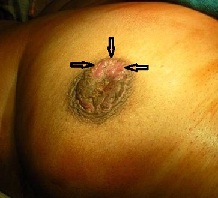
Figure 1: Paget’s disease of the nipple showing ulceration of the nipple areola complex marked by the black arrows
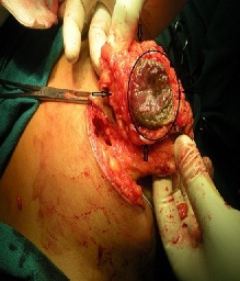
Central quandratectomy was performed which included nipple areolar complex along with rim of normal appearing tissue (Figure 2). Frozen section examination confirmed presence of underlying malignancy (DCIS) with resection margins free of tumor. Final histopathological examination revealed high grade ducal carcinoma in situ with comedo pattern,
receptor studies showed absence of estrogen and progesterone receptors. However HER 2 neu was positive. Postoperatively she underwent whole breast irradiation. Patient has been following up for last 2 years with no clinical or radiological evidence of recurrence.
 s
s
Figure 2: The nipple areola including the lesion marked by the black circle along with a rim of normal breast tissue marked by black arrows removed.
Discussion
The eczematous presentation of Paget’s disease of nipple is the most misleading feature of this disease. Such patients are usually treated by the primary care physician mistakenly for eczema. This may lead to progression of disease eventually resulting into an associated lump with enlarged axillary lymph nodes. Velpeau first described the eczematous lesions of Paget’s disease of nipple in 1856 but it was Sir James Paget who first described the association of Paget’s disease of breast with underlying breast cancer in 1874, and accordingly this condition was named after him [1]. Paget’s disease of the breast is an uncommon presentation of breast with a reported incidence of 3% [2, 3, 4]. Though it is more frequently found in females, it is seen even in males in whom it has a very poor prognosis [3]. The disease to start with is in the form of an eczema which leads to scaling and encrustation. This crust sloughs off resulting in total destruction of nipple areolar complex. This may be associated with symptoms of itching, nipple ulceration and bleeding [3, 4]. Persistent unilateral eczematous dermatitis does not differentiate Paget’s from eczema which may be bilateral but may not persist for long nor does eczema cause destruction of the nipple areola complex. The incidence of underlying cancer in Paget’s disease of nipple is approximately 90% [4, 5, 6, 7]. 50% to 60% of patients with Paget’s disease present with a palpable mass in their breast [1, 3]. Most cases are diagnosed in women in their sixth or seventh decade of life with a mean age at diagnosis reported at 62.6 years [3]. But the age wise incidence is similar to that of the typical breast cancer and can affect younger patients or very old ladies [4]. The associated cancer may not necessarily be located adjacent to the nipple areola complex. The nature of the associated cancer may either be DCIS (10%) or invasive cancer (90%) [5]. Conditions closely simulating Paget’s disease of the nipple include eczema, erosive adenomatosis of nipple, Pagetoid basel cell carcinoma, Bowen’s disease and melanoma [5, 6]. Hence full thickness tissue biopsy from the edge of the nipple is essential to arrive at a pathological diagnosis. Histologically Paget’s disease is characterized by epidermal invasion by malignant glandular cells, which are large, foamy cells that may contain mucin [5, 6]. On staining with hematoxylin and eosin, these cells typically show pale cytoplasm, eccentric and hyperchromatic nuclei and are called as Paget cells which are found throughout the epidermis [6]. Histological diagnosis should be followed by a complete evaluation of the breast for presence of multicentricity [6]. The presence of palpable mass is usually associated with invasive cancer [3, 4, 6]. In women without a palpable mass the lesion is usually a DCIS as is the case presented. Palpable tumor confirmed mammographically in 90% of the cases is usually an invasive carcinoma, and in more than 50% of such cases axillary involvement is present which worsens the prognosis [7 ]. MRI is an excellent investigation which helps in the diagnosis [9, 10]. CT scan of the breast is not as sensitive as MRI especially in smaller lesions. There is paucity of literature to support the use of routine CT scanning in suspected Paget’s disease. Controversy still persists about origin of Paget cells around nipple. Whether these cells arise from the ductal system of the breast or whether these cells are a result of in situ malignant transformation continues to be a matter for debate. Various theories have been proposed for the pathogenesis of Paget’s disease. These are the epidermotropic theory and the in situ malignant transformation theory [1 2]. The first theory states that changes typical of Paget’s disease arise in the ductal cells primarily and later on spread along the basement membrane through the lactiferous sinuses to the nipple. This theory is accepted by virtue of the fact that most patients with Paget’s disease have underlying breast cancer, and the cells from the nipple are histologically similar to the associated invasive carcinoma. The in situ malignant transformation theory on the other hand proposes that Paget’s disease primarily originates in the epidermal cells of the nipple by malignant transformation of keratinocytes and is not associated with any coexisting neoplastic process in the affected breast. A proper full thickness tissue biopsy from the edge of the lesion accompanied with cytological examination of exudates is therefore helpful in not only ascertaining the histology but also helps in evaluation for hormonal receptors [2, 3, 4]. The carcinoma is usually poorly differentiated with absence of receptors ER and PgR but over expresses HER2 neu, as in the case presented [6]. Prognosis of Paget’s disease depends entirely upon the biological features of the underlying carcinoma and is not affected by the presence or absence of DCIS involving the nipple when matched for other prognostic factors [6].
The best surgical option for the Paget’s disease of the breast continues to pose a dilemma. A study of literature fails to reveal clarity on the various surgical options. Traditionally a grossly invasive tumor of the breast necessitates a modified radical mastectomy with axillary clearance. However newer options have evolved over a period of time taking in to consideration a variety of diverse studies comparing survival statistics in patients undergoing radical surgery verses conservative surgery. Paget’s disease of breast therefore happens to be in the twilight zone. The traditional concept supports a radical approach even to Paget’s disease of breast. However two important risk factors require serious consideration prior to determination of surgical options, which include presence of invasive carcinoma and a palpable lump at presentation [2, 3]. A conservative surgical option in the form of breast conserving surgery must be considered in DCIS only. However if there is an invasive cancer with an accompanying mass it would be a safe practice to go for radical surgery. Conservative surgery for Paget’s disease comprises of complete resection of nipple areolar complex ensuring tumor free margins confirmed intraoperatively by frozen section. In such cases an axillary dissection may not be necessary which is based on the assumption that the chances of invasiveness are negligible when the basement membrane is intact. However if the basement is breached while evaluation of such specimen it would be prudent to consider an axillary node sampling. Sentinel lymph node biopsy may be an alternative option to such cases. Various studies have supported high degree of accuracy in identification of sentinel lymph node in patients with Paget’s disease of breast [1, 7]. Sentinel lymph node biopsy should be done in centers where it is a routine practice. However in centers where expertise is not available it is safer to go for an axillary lymph node sampling. Though breast cancer now a day in any form is considered a systemic disease yet good surgical intervention undoubtedly helps in improving disease free survival rate. Extensive literature from all over the world may lead to confusion while deciding the best therapeutic approach to Paget’s disease of the breast. In the Indian scenario where follow up is guided by literacy and the socioeconomic status, one should not consider a conservative surgical approach in breast malignancy as this may adversely affect the survival of the patient. Therefore conservative surgical approach for Paget’s disease of the breast should be an urban phenomenon restricted to specialist centers only. However for patients from rural or socioeconomically underprivileged classes the traditional oncosurgical approach of modified radical mastectomy with axillary clearance is undoubtedly the safest option.
Having offered the best surgical option to the patient, adjuvant therapy is decided by the merits of the case. For a localized DCIS undergoing breast conserving surgery irradiation following surgery should be considered [11, 12]. The role of chemotherapy and HER2 neu receptor blocking agents in the management of DCIS after surgery is not substantiated [2, 3, 12, 13]. But hormonal therapy should be considered even in case of DCIS [2 3]. In cases wherein there is evidence of invasiveness with positive lymph node involvement then adjuvant systemic therapy along with irradiation is indicated. Invasive form of Paget’s disease associated with lump and lymph node enlargement should be treated on the same guide lines as for a classical breast cancer.
Conclusions
A unilateral eczematous nipple lesion needs to be evaluated promptly by way of a breast biopsy.
MRI plays a very important role in diagnosing the underlying lump as compared with other imaging modalities.
Paget’s disease accompanied with an invasive tumor with or without lymph node involvement decides best the surgical option and need for adjuvant therapy.
Conflict of Interest
The authors declare that there are no conflict of interests.
Authors’ Contribution
KV: Contributed to the preparation and editing of the
manuscript.
RC: Edited the photographs and carried out a literature
search.
JP: Contributed to the review of literature and preparation
of manuscript.
MI: Contributed to editing of the manuscript.
AM: Contributed to preparing the photographs and preparing
the manuscript.
All authors read and approved final manuscript for publication.
Funding
None declared
Ethical Considerations
Written informed consent was obtained from the patient for publication of this case report.
References
[1].Osther PJ, Balslev E, Blichert-Toft M. Paget’s disease of the nipple. A continuing enigma. Acta Chir Scand.1990 May; 156(5): 343-52 [Pubmed].
[2].Dalberg K, Hellborg H, Warnberg F. Paget’s disease of the nipple in apopulation based cohort. Breast Cancer Res Treat. 2008 Sep; 111(2): 313-9 [Pubmed].
[3].Sakorafas GH, Blanchord K, sarr MG, Farley DR. Paget’s disease of the breast. Cancer treat Rev. 2001 Feb; 27(1): 9-18 [Pubmed].
[4].Sakorafas GH, Blanchard DK, Sarr MG, Farley DR. Paget’s disease of the breast: a clinical perspective. Langenbecks disease of the breast. 2001 Nov; 386(6): 444-50 [Pubmed].
[5].Lev Schelouch D, Sperber F, Gat A, Klausner J, Gutman M. Paget’s disease of the breast. Harefuah. 2003 Jun; 142(6): 433-7 [Pubmed].
[6].Paone JF, Baker RR. Pathogenesis and treatment of Paget’s disease of the breast. Cancer. 1981 Aug; 48(3): 825-9 [Pubmed].
[7].Chaudhary MA, Millis RR, Lane EB, Miller NA. Paget’s disease of the nipple: a ten year review including clinical, pathological and immunohistochemical findings. Breast Cancer Res Treat. 1986; 8(2):139-46 [Pubmed].
[8].Seetharam S, Fentiman IS. Paget’s disease of the nipple. Women’s Health (Lond Engl) 2009 Jul; 5 (4): 397-402.
[9].Gunhan Bilgen I, Oklay A. Paget’s disease of the breast: clinical, mammographic, sonographic and pathologic findings in 52 cases. Eur J Radiol. 2006 Nov; 60(2): 256-63.[Pubmed].
[10].Capobianco G, Spaliviero B, Dessole S, Cherchi PL, Marras V, Ambrosini G, Meloni F, Meloni GB. Paget’s disease of the nipple diagnosed by MRI. Arch Gynecol Obstet. 2006 Aug; 274(5): 316-8. [Pubmed].
[11].Rickard MT, Selopranoto US. Paget’s disease of the breast: What the radiologist may expect to find. Austral radiol. 1995 Feb; 39(1): 27-30 [Pubmed].
[12].Jamali FR, Ricci A Jr, Deckars PJ. Paget’s disease of the nipple areola complex. Surg Clin North Am. 1996 Apr; 76(2): 365-81 [Pubmed].
[13].Inwang ER, Fentiman IS. Paget’s disease of the nipple. Br J Hosp Med. 1990 Dec; 44(6): 392-5.[Pubmed].

