Case Report

Traumatic vulvar hematoma at term pregnancy
Vani Aditya and Rashmi Malik
Department of obstetrics and gynecology B. R. D. Medical College, Gorakhpur, India and Department of obstetrics and gynecology U.C.M.S. & G.T.B. Hospital, Delhi, India
This is an Open Access article distributed under the terms of the Creative Commons Attribution License (http://creativecommons.org/licenses/by/3.0), which permits unrestricted use, distribution, and reproduction in any medium, provided the original work is properly cited.
Abstract
Background
The overall incidence of trauma during pregnancy is about 5 to 7%. Among the different injuries sustained, trauma to the vulva has not been reported. Even outside obstetrics, traumatic vulvar hematomas are rare. The incidence is not known and literature is limited to the reported cases. The present case addresses the issues related to the management of non obstetric traumatic vulvar hematoma in an obstetric patient at term gestation.
Case Presentation
A gravida 5 woman presented at term gestation with a traumatic vulvar hematoma which was successfully managed by emergency incision and drainage. An uneventful vaginal delivery ensued thereafter.
Keywords
hematoma, pregnancy, term, trauma, vulvar
Introduction
Vulvar hematomas are rare and usually seen in obstetric population following repair of birth related injuries like episiotomies and lacerations. Outside the obstetric population, vulvar hematomas are rarer and usually follow blunt trauma. A wide range of objects like bicycle cross bars, playing toys and boot bindings on snowboards have been associated with saddle injuries [1,2,3]. Spontaneous vulvar hematomas are also known and cases have been reported both in obstetric and non obstetric population [4, 5, 6]. Vulvar hematomas in women with advanced pregnancy or in labor may pose them to risk of complications like rupture or obstruction of labor. Moreover, there is only scant literature on the management of these women in regard to the route of delivery following hematoma repair. The present case addresses the perplexities in the management of such a case of traumatic vulvar hematoma in a pregnant woman at term gestation.
Case Report
A 27 year old, G5 P2 woman presented to accident and emergency unit at 374/7 weeks’ gestation with complaints of intermittent abdominal pain, painful vulvar swelling and retention of urine since nine hours. There was history of fall on abdomen when she straddled a bucket while bathing. Fetal movements were adequate and vaginal bleeding was minimal. Four hours later, when the hematoma was observed to have grown in size, obstetrician’s opinion was seeked. Vitals were stable and mild pallor was noted. The uterus was non-tender and relaxed. Fetal heart was regular. Local examination revealed a 9x5x4 cm3 hematoma involving mainly the left labia minora and to some extent the right labia minora (Fig.1).
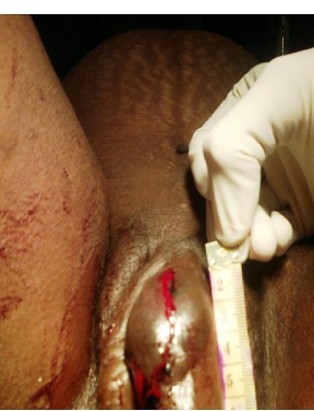
Figure 1: 9cm x 5cm x 4cm size hematoma involving left labia minora.
Both the labia majora were unremarkable. Non stress test was reactive. Ultrasound revealed a single live fetus in cephalic presentation with estimated weight of 2.9 kg. Placenta was located in fundus with no evidence of retroplacental clots. Platelet count, prothrombin time and activated partial thromboplastin tests were normal. Examination and drainage was hence planned under spinal anesthesia. Skin was found to be lacerated and devitalized in medial side of left labia minora with the hematoma extending beneath the clitoris to the right side. Clear stream of urine drained on catheterizing the bladder. Grade III vulvar injury, (AAST, Vulval Injury Scale), was recorded [7]. On vaginal examination, cervix was 2cm dilated and 50% effaced, soft and central. Vertex was at -3 station, membranes were present and pelvis was adequate.
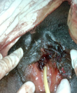
Figure 2.jpg: Restoration of anatomy after hematoma repair.
Hematoma was now evacuated through incision at the site of laceration in the left labia minora. Clots from right side were also milked through the same incision. In all about 600 - 800 ml of clots were removed. An active bleeder was present which was secured. Small necrotic skin tags were removed and dead space obliterated. Incision was closed with restoration of local anatomy (Figure 2). Satisfactory healing by primary intention was seen by day5. For the next two weeks the patient did not go into labor. At 40 weeks, when Bishop Score was even better, cervix was sweeped to induce labor. A 2.8 kg weight healthy baby was delivered vaginally with minimal trauma at the site of previous repair and a small 2°perineal tear (Fig 3).
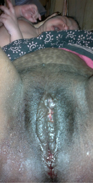
Figure 3.jpg: Uncomplicated vaginal delivery after 2 weeks
Discussion
Blunt trauma to the vulva is rare. Perineum is a highly protected region due to reflex adduction of thighs in the face of impending trauma. Rich vascular supply and its anatomical location however make it susceptible to frontal injuries. The soft tissue of vulva is compressed between the object and the underlying pubic bone leading to laceration and hematoma formation. During pregnancy, physiological increase in vascularity of reproductive organs, venous insufficiency and pathological alterations in coagulation accentuate hematoma formation. Though trivial, vulvar hematomas are important particularly in women who present close to delivery as labor may be complicated with rupture of hematoma or obstruction.
Outside pregnancy, the incidence of vulvar hematomas is not known and literature is limited to several reported cases. Due to rarity, there are no general guidelines for the management. Benrubi et al., found that conservatively managed patients had a longer hospital stay and more interventions subsequently if the product of longitudinal and transverse diameters of hematoma is ≥ 15 cm [8]. In another study by Kanai et al, hematomas with the long diameter > 5 cm were treated surgically and all the cases healed without any sequelae [3]. Evacuation of clots prevents infection and tissue necrosis.
However, a retrospective study by Propst et al observed that, in the absence of acute hematoma expansion, conservative management was often successful [9]. Selective arterial embolization of bleeding vessel has also been found to be successful in management of puerperal and non puerperal vulvar hematomas [2, 6].
The literature on the management of vulvar hematoma during pregnancy is limited just to few case reports. One case of rapidly expanding spontaneous intrapartum hematoma was met with catastrophic outcome due to rupture at the time of delivery [4] . In another case, drainage of spontaneous antepartum hematoma at 35 weeks’ gestation was met with successful outcome and uneventful vaginal delivery four weeks later [5]. In a case of Gravida2, Para1 woman, the authors were skeptical regarding the route of delivery. The woman presented at 32 weeks with giant vulvar hematoma. Though the hematoma was evacuated emergently, abdominal route was chosen for delivery at term gestation because of the possible risk of rebleeding of the vessels in the hematoma cavity during vaginal delivery [10]. In the present case also, the hematoma, presenting in a patient of term pregnancy, was gradually expanding and hence promptly evacuated anticipating complications. Dilemma however existed about the route of delivery. As the patient was expected to progress into active labor, trauma to the site of repair at the time of vaginal delivery was apprehended. Exposure to repeat anesthesia, in case cesarean was indicated later on for obstetric or trauma related indications, was also speculated. Yet, the reasons were not considered sufficient enough to indicate a caesarean delivery at the time of evacuation of hematoma. Considering her multiparous status, average weight of the baby and a reactive NST, the patient was allowed to go into spontaneous labor. Luckily she had a successful vaginal delivery with minimal trauma to the repair site two weeks later. However, some queries remain unanswered. As any wound can resist normal stress such as tension or twisting only after 15 to 20 days i.e., after collagen deposition in the reconstructive phase of wound healing, what would have happened to the wound involving the periclitoral region and labia minora if the patient went immediately into labor? Should caesarean section have been done to prevent wound disruption or rebleeding? to conclude, in women facing imminent delivery, vulvar hematomas may not be as trivial as indicated by trauma scoring. Labor may be complicated by rupture of hematoma or obstruction. With aggressive surgical management, a favorable fetal as well as maternal outcome may be possible. The trauma scoring systems for vulvar injuries should be modified accordingly so that a prompt treatment is given when such patients present to the emergency department. Perplexities regarding the route of delivery, for women who are in active labor following repair of vulvar hematoma, need to be answered and reporting of more such cases will enable us to make evidence based decision about the route of delivery.
Learning Points
What is known?
Outside pregnancy, even large hematomas have been managed conservatively.
What we know now?
a) In pregnant women with term gestation, moderate to large vulvar hematomas should always be treated emergently by aggressive surgical management.
b) Women may safely be allowed to go for vaginal birth if onset of labor is two to three weeks after the repair of vulvar hematoma.
What we need to know?
More cases need to be reported to make a consensus on the route of delivery for women who present with vulvar hematoma when they are in active labor.
Conclusion
In women facing imminent delivery, vulvar hematomas may not be as trivial as indicated by trauma scoring. Labor may get obstructed or be complicated by rupture of hematoma. The scoring systems for vulvar injuries should therefore also consider pregnancy into account so that a proper timely treatment is given when such patients present to the emergency department. An aggressive surgical management of hematoma should be done to achieve a favorable fetal and maternal outcome. For those who are in active labor immediately following hematoma repair, whether a vaginal delivery is safe or a caesarean delivery should be done to prevent wound disruption needs to be answered. Reporting of more such cases will enable to make evidence based decision about the route of delivery.
conflict of interest
There is no conflict of interests
Funding
None
References
[1].Virgili A, Bianchi A, Mollica G, Corazza M. Serious hematoma of the vulva from a bicycle accident: A case report. J Reprod Med 2000;45(8):662 [pubmed][pubmed]
[2].Kunishima K, Takao H, Kato N et al. Transarterial embolization of a nonpuerperal traumatic vulvar hematoma. Radiat Med. 2008;26(3):168 [pubmed][pubmed]
[3].Kanai M, Osada R, Maruyama KI. Warning from Nagano: Increase of vulvar hematoma and/or lacerated injury caused by snowboarding. J Trauma 2001;50(2):328 [pubmed][pubmed]
[4].Joy SD, Huddleston JF, McCarthy R. Explosion of a vulvar hematoma during spontaneous vaginal delivery: A case report. J Reprod Med.2001;46(9):856 [pubmed][pubmed]
[5].Nelson EL, Parker AN, Dudley EJ. Spontaneous vulvar hematoma during pregnancy: A Case Report. J Reprod Med.2012; 57(1-2):74 [pubmed][pubmed]
[6].Egan E, Dundee P, Lawrentschuk N. Vulvar hematoma secondary to spontaneous rupture of the internal iliac artery: clinical review. Am J Obstet Gynecol. 2009;200:e17 [pubmed][pubmed]
[7].Moore EE, Jurkovich GJ, Knudson MM et al. Organ injury scaling. VI: Extrahepatic biliary, esophagus, stomach, vulva, vagina, uterus (nonpregnant), uterus (pregnant), fallopian tube, and ovary. J Trauma. 1995;39(6):1069 [pubmed][pubmed]
[8].Benrubi G, Neuman C, Nuss RC et al. Vulvar and vaginal hematomas: a retrospective study of conservative versus operative management. South Med J.1987; 80(8):991 [pubmed][pubmed]
[9].Propst AM, Thorp JM Jr. Traumatic vulvar hematomas: Conservative versus surgical management. South Med J.1998; 91(2):144 [pubmed][pubmed]
10].Hacivelioglu S, Haydardedeoglu B, Simsek E, Cok T. Giant vulvar hematoma during pregnancy after sexual intercourse:A case report East J Med 2012;17:94

