Original Article

Persistent correctable urinary dribbling in a female child: A cases series
*Pranaya Panigrahi,*Rakesh Kumar, *, Quamaruzzama Ansari, *Vaibhav Pandey
- 1Pediatric surgery, MKCG MCH, Brahmapur, Odisha, India
- 2Department of Pediatric Surgery, IGIMS, Patna, Bihar, India
- 3Department of General Surgery, Career Institute of Medical Sciences, Lucknow, U.P.
- 4Department of Pediatric surgery, Institute of Medical Sciences, Banaras Hindu University, Varanasi, U.P., India
- Submitted: Thursday, July 9, 2020
- Accepted: : Monday, May 18, 2020
- Published: Saturday, August 1, 2020
This is an Open Access article distributed under the terms of the Creative Commons Attribution License (http://creativecommons.org/licenses/by/4.0), which permits unrestricted use, distribution, and reproduction in any medium, provided the original work is properly cited
Abstract
Introduction
Persistent urinary dribbling in a female child is not uncommon in clinical practice. Many female babies either not adequately toilet trained or having congenital pathology present to pediatric urologist late. Reason being parents delaying to report it considering continence attaining age of their child late or a part of social stigma.
Methods
This case series reports three females of pediatric age group with correctable condition presenting as dribbling owed to ectopic ureter in different locations. All cases were managed surgically after diagnosis and assessment of renal function. Cystoscopy with
genitoscopy whenever needed was done to ascertain the anomaly. Open or Laparoscopic intravesical reimplantation with or without ligation of ectopic ureter was performed in all cases. They were advised for follow-up and renal function status was monitored.
Results
All cases were managed by open or minimal invasive approach following diagnostic cystoscopy. All cases were doing well in post operative period and follow-up. They are in follow-up with us with well preserved renal function on renal scan.
Conclusion
Persistent dribbling is to be addressed with all available options and should be managed in pediatric urology centers in order to ascertain renal status and surgical corrections if any.
Keywords
UTI, re-implantation, ectopic ureter, Ureteric duplication, VUR, Laparoscopy
Introduction
Chronic urinary discharge/dribbling during infancy is a common concern although there is usually no underlying urinary tract abnormality in majority of cases. In females, urinary tract infection (UTI), chronic constipation with improper toilet training may be listed as common causes. In contrast, urinary incontinence is also the main presenting feature of an ectopic ureter, especially in females because of urinary tract infection (UTI) or congenital obstructive uropathy. An ectopic ureter can present as a prenatal diagnosis in both sexes [1]. It occurs in 1 in 2000 patients, with the female to male ratio being 6:1 [2].
Bilateral complete renal duplication with ureteral ectopia is an unusual condition, occurring most frequently in females. There is no known statistics regarding the incidence, because it is mainly an incidental finding [2]. Clinical complications arise from congestion, reflux, or ectopic openings, which lead to hydronephrosis, inflammation, and incontinence [3]. Some cases are diagnosed as a consequence of chronic urinary dribbling or persistent UTIs in duplex kidneys with dysplastic upper renal pole moiety throughout childhood. Due to inadequate access to adequate health care, or due to milder symptoms over years, some patients may develop symptoms during adulthood.
This case series compiles very unusual presentations of persistent urinary dribbling. Also illustrates the difficulties of diagnosing and treating this debilitating condition versus several conditions that may occur with these symptoms e.g voiding dysfunction, overly active bladder, and unilateral ectopic ureter [4]. In addition, upgraded difficulty to diagnose and treat young patients with urinary incontinence after a proper toilet training or repeated UTIs.
Case description
Case 1: Bilateral complete duplication with ectopic ureter in vagina.
A 5-year-old girl child present to us in outpatient department with history of urinary
dribbling since birth. There was no history of recurrent UTI. She was constantly wet but had normal voiding habits. The patient had continuous low volume urine leakage requiring 4–5 daily pads. The parents could not specify whether there was any connection with standing, coughing, or
sternous effort and she had no urge to void. The systemic and genital examination was normal. Ultrasonography showed possibility of bilateral duplex system. A voiding cystourethrogram (VCUG) was performed and a vesicoureteral reflux (VUR) grade III
was found on the right side. On Cystoscopy and genitoscopy, we found that there were two ectopic openings
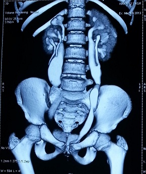
seen in the vaginal wall one on each side. A contrast-enhanced computed tomography (CT) of the abdomen and pelvis was performed to visualize the entire urinary tract. It showed bilateral complete duplication with bilateral ectopic ureters in vagina. [Figure 1] An exploration with ligation of ectopic ureters along with Ureteric Re-implant on right side was performed.[Figure 2]
.jpg)
Fig 1 :CECT showing bilateral duplication with ectopic opening in vagina
.jpg)
Fig 2: Bilateral intravesical ureteric reimplantation
Case 2: Unilateral ectopic in vagina without duplication
A 3 year old girl child presentd to our outpatient department with history of urinary
dribbling since birth. She was constantly wet but had normal voiding habits and had no history of recurrent UTI. The systemic and genital examination was normal. Ultrasonography showed right sided mild hydroureteronephrosis. VCUG was normal. We performed cystoscopy which showed ectopic opening in vagina and only left Ureteric orifice in bladder. Diagnosis of right ectopic ureter without duplication was made. Renal scan showed good renal function on both sides. Exploration and ureteric reimplantation was performed of the right ureter.
Cases 3: Unilateral duplication with ectopic below the bladder neck with bilateral VUR
A 6-year-old girl child presented with history of recurrent episodes of UTI. On examination there was excoriation and parents gave history of continuous dribbling of urine. Rest of systemic and genital examination was normal. Ultrasonography showed right sided hydroureteronephrosis with possibility of duplication. VCUG showed bilateral Grade V VUR [Figure 3]. We performed Cystoscopy which showed ectopic ureteric opening just below the bladder neck [Figure 4]. In bladder there were two ureteric orifices with enlarged refluxing left orifice. A CECT of whole renal system was performed which reveled duplicated right sided system with ectopic opening. A laparoscopic ureteric reimplantation was performed.

Fig 3: VCUG showing bilateral Grade V VUR

Fig 4: Cystoscopy showing ectopic ureteric opening at the bladder neck
Discussion
In an autopsy series, the incidence of complete or incomplete urinary tract duplications in the urinary system was reported to be 0.7% and in clinical series between 2% and 4% [5]. In women it is twice as common as in men [6]. Symptoms of the patient depends on the ectopic ureter insertion site and this varies between girls and boys [7]. Males typically may not have urinary incontinence due to the insertion of the ureter over the external urinary sphincter, but may have antenatal hydronephrosis or UTI. Some cases are diagnosed from persistent urinary dribbling or repeated UTIs during childhood.
Three times as much as complete duplication is seen as incomplete
duplication. Complete duplications are uncommon, present in <0.1% of the population, and are more prevalent in females [2]. For these, only 25% are stated to be bilateral [2].
Ectopic ureter is characterized as an anomalous opening in the trigone of the ureter orifice outside its usual location. This condition is more frequently seen in females and is correlated with the duplex collection system at 80% [6]. Weigert Meyer law is complied with in cases of complete ureteral duplication [8]. Compared with the ureter of the lower pole moiety [8], the ectopic ureter coming from upper moiety is inferior and medial. Furthermore, as the lower pole ureter makes a shorter path into the bladder, it is more resistant to reflux, while the upper pole ureter's anomalous penetration renders this moiety more susceptible to obstruction [9]. In rural areas or poor urban areas, the manifestation of congenital abnormalities in late life is more likely to be seen. [10].
The presenting symptoms depend on the ectopic ureter insertion site and this varies between boys and girls [7]. Males do not usually suffer urinary incontinence because ureter is inserted just above the external urinary sphincter. Such as in our cases, many females present with urinary dribbling, as ectopic ureter insertion is distal to the external urinary sphincter. Generally affected females have low volume leakage or spotting incontinence with intact voiding patterns [11].
For females, bladder neck and upper urethra (33%), vestibule between urethra and vaginal opening (33%), vagina (25%), and less commonly the cervix or uterus (< 5%) are the most common ectopic ureter insertion sites [7]. Those female with a ureteral insertion on or above the neck of the bladder would be continent [1].
Renal ultrasound and excretory urography will never completely detect the ectopic insertion of a ureter which does not provide sufficient data on the exact anatomy and association of ureter, bladder and vagina [11]. Also the preferred option for depicting or ruling out an ectopic ureter should be the contrast-enhanced CT or MR urography [11]. Additionally, associated renal dysplasia, congenital heart disease, spinal cord malformations, anorectal malformations can be associated with an ectopic ureter which was absent in our cases.
Children with symptoms are better treated with surgery with attempts to overcome incontinence, avoid further complications, maintain renal function, and decrease UTIs [11]. In patients with preserved renal function, ureteral laparoscopic ligation (clipping) and ureteral reimplantation is warranted. Non-functioning pole hemi-nephrectomy with or without re-implantation in the duplex system after renal function scan is mandated in cases of duplex system with ectopic ureters. [12].
Conclusion
Ectopic ureter should be a kept as a differential
diagnosis in cases of persistent wetting and recurrent urinary tract infections after excluding other common subtle correctable etiologies. Endoscopic examination certified the location of the ectopic ureter orifice in our cases mandating to be kept in protocol for assessment in cases of female dribbling. Laparoscopic approach could be helpful as excellent visualization during laparoscopy helps to delineate ureter’s course, better preservation of renal pedicle especially in management of atrophic renal moieties. Symptoms, renal function, patient's age and life quality will be the ones deciding the management of an ectopic ureter.
Conflict of Interest
None Declared
Author’s contribution
PP:
Conceived and designed the study, analysis interpretation and preparation
of manuscript.
RK:
Conceived and designed the study, collected the data and analyzed it.
QA:
Consent, counselling, data analysis, interpretation and other.
VP:
Conceived and designed the study, analysis interpretation
All authors have read the final manuscript and approve it for
publication
Ethical Statement and Consent
The written informed consent was obtained from the father of the patients and is available with authors
Funding
None
Acknowledgements
None
References
[1]. Baskin Laurence S. Ectopic Ureter. Up
to date (2016) Available online at:
http://www.uptodate.com/contents/ectopic-ureter [Last accessed May 21, 2020]
[2]. Mittal MK, Sureka B, Gupta P, Mittal A. Bilateral duplex system with overlooked dysplastic moiety: A rare cause of incontinence. J Mahatma Gandhi Inst Med Sci. 2014; 19:67-69.
[Full
Text]
[3]. Kate DR, Shinde B (2015) Duplex kidney-an anatomical and clinical insight: IOSR. Journal of Dental and Medical Sciences. 2015; 14-17.
[4]Hanson GR, Gatti JM, Gittes GK, Murphy JP. Diagnosis of ectopic ureter as a cause of urinary incontinence. J Pediatr Urol. (2007) 3:53–7. [PubMed]
[5]. Cassell AK, Traoré A, Jalloh M, Ndoye M, Diallo A, Labou I, et al. Bilateral Ureteral Duplication and Right Ectopic Ureter Presenting with Incontinence: A Case Report. Med Sur Urol 2019; 8:216.
[Full
Text]
[6]. Schulman CC. The Ureter; in O’Donnell B, KoffS A. Pediatric Urology.
Edited by B O'Donnel, SA Koff. Oxford, Butterworth Heineman, 1997; 397-418.
[7]. Nzenza TC, Rice G, Kinnear N, Hennessey D. An interesting case of lifelong urinary incontinence. Austin J Urol. (2016) 3:1048.
[Full
Text]
[8]. Fufezan O, Tatar S, Dee AN, Cramariue R, Asavoaie C, Coşarcă M. Large spectrum of complete urinary collecting system duplication exemplifid by cases. Pictorial essay. Med Ultrason. 2013; 15:315-320.[Pubmed]
[9]Callahan MJ. The drooping lily sign. Radiology. 2001;219:226-228.[Pubmed]
[10].Ohmann EL, Borofsky MS, Han JS. Unusual presentation of ectopic insertion of duplicated collecting system in an adult male. Urology. 2013; 81:e36-e37.[pubMed]
[11]. Duicu C, Kiss E, Simu I, Aldea C. A Rare Case of Double-System with Ectopic Ureteral Openings Into Vagina. Front Pediatr. 2018;6: 176.[PubMed] [PMC full Text]
[12]. Romao RL, Figueroa V, Salle JL, Koyle MA, Bägli DJ, Lorenzo AJ. Laparoscopic ureteral ligation (clipping): a novel, simple procedure for pediatric urinary incontinence due to ectopic ureters associated with non-functioning upper pole renal moieties. J Pediatr Urol. 2014; 10:1089-1094.[Pubmed]

