Case Report

Reconstruction in a Patient with Giant Cell Granuloma
*Dr. Deepak Kumar Gupta,*Dr. Sudhir Kumar Majhi
- *Department of ENT, VMMC & Safdarjung Hospital
- Submitted 30th January 2018
- Accepted 23rd February 2018;
- Published 30th March 2018
This is an Open Access article distributed under the terms of the Creative Commons Attribution License (http://creativecommons.org/licenses/by/4.0), which permits unrestricted use, distribution, and reproduction in any medium, provided the original work is properly cited
Abstract
Giant Cell Granuloma is a rare benign disease. We present a case of Giant Cell Granuloma involving mandible in a 7 year old boy. Rarity of the disease, difficulty in diagnosis along with surgical treatment including reconstruction employed in this case is discussed.
Keywords
Mandible, giant cell granuloma, reconstruction.
Introduction
Giant Cell Granuloma constitutes 10% of all the benign tumors of jaw. It usually affects children and young adults in children with male preponderance. It is more commonly located in the mandible than in maxillary bone. It is often difficult to differentiate it from aneurysmal bone cyst. Reconstruction after excision of these tumors is challenging. Autograft using rib has excellent success rate both functionally as well as cosmetically.
Case Report
A seven year old male child reported in ENT OPD of VMMC & Safdarjung hospital with a gradual increasing swelling over the right side of jaw after trauma for the past three month. He also had difficulty in opening the month and chewing the food.
On examination the swelling extended superiorly to the right ear lobule, inferiorly at the level of hyoid bone, posteriorly to the anterior border of SCM and anteriorly to the angle of mouth. Swelling was firm in consistency, tender, temperature was not raised and was fixed to the underlying hemimandible. Skin over the swelling was normal (fig 1).Oral cavity proper was within normal limit. The patient was admitted and Orthopantomogram was done (fig 2), which revealed an expansile radiolucent osteolytic lesion involving body and ascending ramus of right hemi mandible. CT scan reveals unilocular cyst without any fluid filled level (fig 3). FNAC revealed possibility of aneurysmal bone cyst. In view of the above finding a differential diagnosis of aneurysmal bone cyst and GCG was made. One stage right hemimandibulectomy and mandibular reconstruction was planned. Patient underwent right hemimandibulectomy (bony swelling with coronoid process, condoyle and ramous of mandible up to mental foramen) (fig 4).
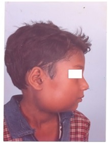

Fig-1:- Pre-operative photograph showing the swelling in right hemimandible

Fig-2:- OPG showing expensile radiolucent osteolytic lesion involving right body and ascending ramus

Fig-3:-CT scan showing unilocular cyst without any fluid level.

Fig-4:- Photograph of specimen
Reconstruction of the defect was done using 6th rib costal cartilage from right thoracic region of the patient by a Plastic surgeon (fig 5). One end of the rib was fixed to the remaining stump of hemimandible by stainless steel wiring and the other end placed inside the glenoid fossa. Inter dental wiring was done for one month and during this period patient was put on Ryle's tube feeding. Histopathology examination revealed fibroblastic and giant cells in low power and high power magnification, suggestive of Giant Cell Granuloma (fig 7 & 8). Postoperative edema and swelling subsided gradually. After removal of inter dental wiring patient was put on semi solid diet. Postoperative OPG was done after 6 months which revealed a good take up of rib graft. Follow up of two years does not show any recurrence with an excellent functional & cosmetic result (fig 6).
Fig-7:-HPE showing fibroblastic and giant cells in low power field

Fig-8:-HPE showing fibroblastic and giant cells in high power field

Discussion
GCG was first described by Jaffe (1) as a locally reparative reaction of bone possibly due to an inflammatory response, hemorrhage or local trauma. Etiology is probably due to reactive process in response to intraosseous hemorrhage following trauma. GCG is considered to be more common in the mandible than in the maxilla. The majority of cases occurs in the molar-premolar area and could extend to the ramus (7, 8). Involvement of the condyle is rare (3-5, 9). Some cases are asymptomatic (8); however, the most common presentation of GCRG is a painless expansible mass in the face or the oral cavity (10). Additionally, ≥20% patients experience pain or paresthesia (1, 6, and 11).
Other symptoms may include facial asymmetry, loosening or displacement of teeth, and pathological fractures (12). It can be of two types nonaggressive and aggressive. Aggressive type may result in extensive tissue destruction at later stage. It is difficult to differentiate it from aneurysmal bone cyst on the basis of FNAC and radiology. FNAC is easy simple and quick method for pre-operative evolution for diagnosis. However it has its limitation in definitive diagnosis due to paucity of cells and lack of architectural context of FNA material. Though in our case FNAC shows Aneurysmal bone cyst but on CT scan, it had unerupted tooth, was unilocular and had an absence of fluid level which favors GCG.
The treatment modalities most frequently used are enucleation, curettage alone. Various modalities of surgical treatment including mandibular reconstruction technique have been employed in a child with GCG of mandible. The goals of reconstruction of a mandibular defect involving the body & ascending ramus of mandible are to achieve a stable articulation and regain continuity, as well as restoring facial form and dental occlusion. There is ongoing controversy regarding the best way to reconstruct the condyle, whether to use autogenous tissues or alloplastic materials. However, in our experience, costochondral rib graft remains the preferred method of reconstruction. It has an excellent take up along with cosmetic and functional result. This is mainly because we are able to achieve consistently good results that are shared by others also (15-17). Retrospective analysis of 38 cases of advance disease during 1970 to 2015 revealed treatment by local excision and partial mandibulectomy using iliac bone graft for mandibular defect reconstruction in 9 cases with good results by Tallan EM, Olsen KD, Maccaffrey TV, Unni KK and Lund BA (3). Icon omen TG, Zuuker RM and Philips JH (1) reported mandibular reconstruction in children using the vascularised fibula in 10'consecutive patients with good results.
Wise AJ and Bridbord JW (2) performed mandibular reconstruction with split rib graft. This technique stimulated new bone formation which provided excellent contour to the angle of mandible in adult patients and also resulted in normal mandibular growth in children. Mandibular reconstruction with split rib graft which was done in our case is more promising as new bone formation provided contour to the angle of mandible and normal mandibular growth in children.
Non-surgical approach in the form of Intralesional Corticosteroids, Calcitonin, & Interferon Alfa - 2A. In order to cure or reduce the size of the lesion and thus minimize the need for extensive surgical resection that could result in functional and cosmetic defects are under trial. Radiotherapy should be avoided because of theoretical risk of long-term malignant transformation (13, 14).
Alloplastic replacement of the mandibular body & ramus has advantages of rigid stabilization and no morbidity of donor-site morbidity. However, its applications have other concerns and potential disadvantages. There is an overall 10% complication rate with metallic alloplastic condylar heads including pain, loose plate, limited jaw opening and plate exposures in irradiated patients (18). Moreover, the alloplastic metallic condylar head is very expensive and some have shown that alloplastic prosthesis is not a suitable option for temporomandibular joint reconstruction(s) (19). In our experience, costochondral rib grafts have been used for reconstruction with good success and have remained our preferred method of reconstruction following tumors resection.
Conflict of Interests
The authors declare that there are no conflicts of Interests.
References
[1]Iconomon T G ,Zuker R M and Phillips JH.Mandibular reconstruction in children using the vascularised fibula .J ReconstrMicrosurg. 1999; 15(2): 83-90.[PubMed]
[2]Wise A J and Bridbord J W .Giant cell granuloma of the facial bones. Ann Plast, 1993; 30(6):564-8.[PubMed]
[3]Tallan E M ,Olsen K D ,McCafrey T V ,Unni K K and Lund B A .Advanced giant cell granuloma: a twenty – year study. Otolaryngol Head Neck Surg.1994; 110 (4):413-8.[PubMed]
[4]Jaffe H L. Giant-cell reparative granuloma, traumatic bone cyst, and fibrous (fibro-oseous) dysplasia of the jawbones. Oral Surg Oral Med Oral Pathol. 1953;6(1):159-75.[PubMed]
[5]Shah U A, Shah A K, Kumar S. Giant cell reparative granuloma of the jaw: A case report. Indian J Radiol Imaging. 2006;16(4):677-678.
http://www.ijri.org/text.asp?2006/16/4/677/32297
[6]The InfanteCossío P, Martínez de Fuentes R, Carranza Carranza A, Torres Lagares D, Gutiérrez Pérez JL. Recurrent central giant cell granuloma in the mandible: Surgical treatment and dental implant restoration. Med Oral Patol Oral Cir Bucal. 2007;12:E229-32.[PubMed][Free Full Text
[7]Kaaban L B, Troulis M J, Ebb D, August M, Hornicek F J, Dodson T B. Antiangiogenic therapy with interferon alpha for giant cell lesions of the jaws. J Oral Maxillofac Surg. 2002;60(10):1103-11.[PubMed]
[8]Kaffe I, Ardekian L, Taicher S, Littner MM, Buchner A. Radiologic features of central giant cell granuloma of the jaws. Oral Surg Oral Med Oral Pathol Oral Radiol Endod. 1996;81(6):720-6. [PubMed]
[9]Tasanen A, Von Konow L, Nordling. Central giant-cell lesion in the mandibular condyle. Report of a case. Oral Surg Oral Med Oral Pathol. 1978;45(4) :532-9.[PubMed]
[10Shensa D R, Nasseri S. Central giant cell reparative granuloma of the mandibular condyle. J Oral Surg. 1978;36(8):642-3.[PubMed]
[11]Abu-El-Naaj I, Ardekian L, Liberman R, Peled M. Central giant cell granuloma of the mandibular condyle: A rare presentation.J Oral Maxillofac Surg. 2002;60:939-41. [PubMed]
[12]Waldron C A, Shafer W G. The central giant cell reparative granuloma of the jaws. An analysis of 38 cases. Am J ClinPathol. 1966;45(4):437-47.[PubMed]
[13]Austin L T, Dahlin D C, Royer R Q. Giant-cell reparative granuloma and related conditions affecting the jawbones. Oral Surg Oral Med Oral Pathol. 1959;12:1285-95.[PubMed]
[14]Ustundag E, Iseri M, Keskin G, Müezzinoğlu B. Central giant cell granuloma. Int J Pediatr Otorhinolaryngol. 2002;65(2):143-6.[PubMed]
[15]Wendt F P, Torriani M A, Gomes A P, de Araujo LM, Torriani DD. Intralesional corticosteroid injection for central giant cell granuloma: An alternative treatment for children. J Dent Child (Chic). 2009; 76(3):229-32.[PubMed]
[16].Carlos R, Sedano H O. Intralesional corticosteroids as an alternative treatment for central giant cell granuloma. Oral Surg Oral Med Oral Pathol Oral RadiolEndod. 2002;93(2):161-6.[Pubmed]
[17]Harris M. Central giant cell granulomas of the jaws regress with calcitonin therapy. Br J Oral Maxillofac Surg. 1993;31(2):89-94.[PubMed]
[18]Hamlin W B, Lund P K. Giant cell tumors of the mandible and facial bones. Arch Otolaryngol 1967;86(6):658-665.[PubMed]
[19]Lindqvist C, Jokinen J, Paukku P, Tasanen A. Adaptation of autogenous costochondral grafts used for temporomandibular joint reconstruction: A long-term clinical and radiologic follow-up. J Oral Maxillofac Surg. 1988;46(6):465-70.[PubMed]

