Case Report

Arteriovenous Malformation of Upper Extremity with Multiple Hemangioma: A Case Report
1 Raju Kumar, 2 Anita Kumari,2Sangeeta Pankaj, 2Jaya Kumari, 2Syed Nazneen,2Anjili Kumari,2Simi Kumari
- 1 1Department of Pediatrics, Indira Gandhi Institute of Medical Sciences, Patna, India
- 2 Department of Gynecological Oncology, Indira Gandhi Institute of Medical Sciences, Patna, India
- Submitted:Tuesday, August 15, 2017
- Accepted: Saturday, October 21, 2017
- Published: Thursday, December 7, 2017
This is an Open Access article distributed under the terms of the Creative Commons Attribution License (http://creativecommons.org/licenses/by/4.0), which permits unrestricted use, distribution, and reproduction in any medium, provided the original work is properly cited
Abstract
Infantile hemangiomas (IH) are the most common vascular tumors of infancy, usually presenting at birth or during the first few months of life and affecting 1-2% of neonates.
Prevalence of arteriovenous malformation is about 1%, out of which 40% AVM appears at birth and most of them are sporadic. We report a rare case of vascular anomalies in an infant which both arteriovenous malformation of left upper extremity and multiple infantile hemangiomas were present.
Key words:
Hemangioma, Arteriovenous malformation, vascular anomalies, Neonate.
Introduction
Pediatric vascular tumour and malformation comprising a broad category of lesions called vascular anomalies and are clinic-pathologically distinct. Infantile haemangiomas (IH) are the most common vascular tumours of infancy, usually presenting at birth or during the first few months of life. In neonatal period, it can be divided into two different classifications: multiple congenital, limited to skin involvement, or as a potentially fatal condition called diffuse neonatal hemangiomatosis (DNH).
Vascular malformations were described as lesions present at birth and composed of vascular channels lined with flat mature endothelium. Vascular malformation divided into low-flow venous malformations (VMs) and high-flow arteriovenous malformations (AVMs). Congenital AVM occur as a result of lack of differentiation of arteries, vein, and the capillaries during vascular development [1]. A persistent communication exists between them resulting in short circulating blood. They always contain a number of abnormal A-V connections with seldom any dramatic cardiovascular effect.
AVMs are a distinct entity characterized by high-flow physiology and an aggressive clinical course. The lesion can be detected at birth in 40% of cases and most commonly occurs in the extremities and pelvis. The diagnosis of presence and type of vascular malformation usually can be made purely on clinical history and physical examination.
Systemic corticosteroids used to be the first-line treatment for IH; however, long term use can lead to serious side effects such as hypertension, adrenal cortical insufficiency, and delay of growth [2].
Case report
A pregnant woman of age 26 years, G-4, P3+0 presented in labour room of Patna Medical College and Hospital, Patna (PMCH) with 39 weeks gestation in active phase of labour. Labour was progressed normally and a female child was delivered vaginally. Baby was pink colored and cried normally at birth. Apgar score at one minute was 8/10 and at 5 minute was 8/10. Her birth description was (i) weight: 2.6 Kg, (ii) HC: 34 cm iii) .On examination there was large lobulated mass in left upper extremity with Multiple violet color papules observed on whole body. Thus, neonate was transferred to neonatal intensive care unit for further management of these lesions.
On physical examination soft spongy lobulated bluish swelling was found on left upper extremity except hand, which was compressible (Figure 1). The size of swelling observed to get decreasing on application of pressure, but on the release of pressure it appears to revert back to its original size. Prominent vessels were present on the surface of the swelling. There was multiple violaceous papules over whole body of the neonate and were blanching (Figure 2).
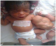
Figure 1 Clinical Photograph

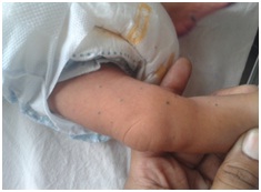
Figure 2 multiple violaceous papules
X-ray of hands and chest were found normal. Ultrasonography showed multiple vascular channels with enlarged draining vein. However, Doppler USG showed arterial and venous signal from vessels in the lesion with arterializations of venous structures. Neonate was hemodynamically stable.
Discussion
Infantile haemangioma, especially when cutaneous, are the most common tumour of infancy, affecting 1 to 2% of neonates. They tend to be more frequent in females and premature baby, and usually present in the first weeks of life, having a characteristic evolution. The haemangioma may occur anywhere on the skin but the head and neck are the most commonly affected sites, followed by the trunk and limbs. In approximately 10 to 20% of babies with haemangioma, the lesions are multiple. Although the exact mechanism for haemangioma development remains unknown, vascular growth factors seem to play a role in the pathogenesis. Proliferation most probably results from an imbalance between positive and negative angiogenic factors expressed by the haemangioma and adjacent normal tissue [3].
Previousely corticosteroid was the main treatment of haemangioma but several studies have confirmed that oral propranolol significantly accelerates the involution rate of IH, even in post-proliferative phases, in children up to 10 years of age.
Schobinger classification outlines the progressive clinical course of an AVM if left untreated. Lesions progress from stage I (quiescence) to stage II (expansion) with increasing pulse and thrill. Stage III is characterized by local destruction associated with pain, ischemia, and necrosis. If left untreated, AVMs can progress to stage IV (decompensation) with high-output cardiac failure.
The diagnosis of a vascular malformation can usually be made correctly on the basis of clinical history and examination alone. Diagnosis is nearly always confirmed with some form of imaging or rarely biopsy. There have been much recent advances in the evaluation of such AVMs with imaging modalities ranging from simple X-ray and sonography to MRI and MRA.
The first investigations usually performed are Ultrasound and Doppler examination which allow immediate differentiation between a low-flow (Vascular or Lymphatic Malformation) and high-flow (AVM) lesion.AVMs on Ultrasonography imaging are usually described as high systolic flow, shunt with spectral broadening within a region of high vessel density due to localized arterial and venous hypertrophy. Although CT scans can provide excellent pictures of the malformations and delineate the extent of involvement but may not always differentiate AVM from venous malformation. MRI and MRA are investigating modalities of choice as they offer many advantages over other imaging modalities.
The patient was diagnosed clinically as having a vascular malformation and Doppler ultrasound helped us describe the type of malformation as a high flow AV malformation. Once diagnosis was made, the next challenge was to choose modality of treatment. The recent years have witnessed a great deal of treatment interventions available for treating AVMs. The commonly used treatment modalities include embolization, sclerotherapy, lasers and surgical excision. According to some surgeons, surgery is effective method of treatment for congenital arteriovenous malformations [4]. A detailed assessment of the nature of the lesion, it’s impact on quality of life, the non-neoplastic nature of the lesion, possible complications and the treatment options available with complications, success and failure rates have to be discussed with the patient’s relative before planning any intervention.
Since before deciding any treatment modality, patient was lost on follow up. We could not plan any intervention and lost opportunity to treat high flow AV malformation and to compare and study our management to other known studies.
Conflict of Interests
The authors declare that there are no conflicts of Interests
Authors’ Contribution
Rk - carried out the experiments and interpreted the results,
AK - carried out the literature search and prepared the draft manuscript,
SP - designed the study and performed the analysis,
JK - conceived the study, participated in design and edited the final manuscript.
SN - conceived the study, participated in design and edited the final manuscript.
AK - conceived the study, participated in design and edited the final manuscript.
SK - conceived the study, participated in design and edited the final manuscript.
All authors read and approved the manuscript.
Ethical Considerations
The written informed consent was obtained from the legal guardian and is available with the authors.
Funding
None Declared
References
[1]Plasencia AR, Santillan A. Embolization and radiosurgery for arteriovenous malformations. Surg Neurol Int. 2012;3(Suppl 2):S90-S104. doi: 10.4103/2152-7806.95420. Epub 2012 Apr 26. [PubMed]
[PMC Full text]
[2]Liu X, Qu X, Zheng J, Zhang L. Effectiveness and Safety of Oral Propranolol versus Other Treatments for Infantile Hemangiomas: A Meta-Analysis. PLoS ONE 2015; 10(9):e0138100.[PubMed]
[PMC Full text]
[3.Lembo S, Balato A, Raimondo A, Donofrio
P, Lembo C, Balato N: A preterm infant with benign neonatal hemangiomatosis and
persistent patent ductusarteriosus: a curious comorbidity. GiornaleItaliano di
Dermatologia e Venereologia 2012, 147(3):321–324 [PubMed]
[4Ozcan AV, Boysan E, Isikli OY, Goksin I. Surgical treatment for a complex congenital arteriovenous malformation of the lower limb. Tex Heart Inst J. 2013;40(5):612-4. [PubMed]
[PMC Full text]

