Case Report

Surgical Treatment of Cyst of the Canal of Nuck: Report of a case.
*Vittorio Lombardo, *Giovanni Di Cara, *Giuseppe Pavone, *Massimo Turrisi, *Giuseppe Cuticone, *Francesco Iaropoli.
- * Division of General Surgery, Papardo-Piemonte Hospital, Contrada Papardo, Messina, Italy
- Submitted: Wednesday, September 6, 2017
- Accepted: Saturday, October 21, 2017
- Published: Monday, November 20, 2017
This is an Open Access article distributed under the terms of the Creative Commons Attribution License (http://creativecommons.org/licenses/by/4.0), which permits unrestricted use, distribution, and reproduction in any medium, provided the original work is properly cited
Abstract
The canal of Nuck is the portion of the processus vaginalis within the inguinal canal in women. A hydrocele of the canal of Nuck is equivalent to an encysted hydrocele of the cord in men. Although the canal of Nuck normally disappears without a trace in the first year of life, it can cause an indirect inguinal hernia or hydrocele of the canal of Nuck when, in rare cases, it does not disappear completely.The cyst formation is likely due to the imbalance of the secretion and absorption of the secretory membrane lining the processus vaginalis. Trauma or infection may cause disruption of lymphatic drainage, which can lead to the imbalance, but in most cases, it is idiopathic. Swelling in the inguinal region of a woman may result from several conditions, including adenopathy, inguinal hernia, cyst, abscess, tumor (lipoma, leiomyoma, sarcoma), or hydrocele of the canal of Nuck. The literature reveals very little about this rare condition in the adult female patient. In this paper, we report a case of hydrocele of the canal of Nuck in a young female. The patient presented with an inguinal swelling on physical examination.The diagnosis was made with ultrasound and magnetic resonance imaging and then confirmed postoperatively and by histopathology. Although rare, a hydrocele of the canal of Nuck has to be included in the differential diagnosis of a groin lump in female patients.
Keywords
Cyst of canal of Nuck, hydrocoele of canal of nuck, excision of cyst of canal of Nuck
Introduction
The round ligament is attached to the uterus and a small evagination of the parietal peritoneum accompanies the round ligament through the inguinal ring into the inguinal canal in the female. This small evagination of the parietal peritoneum is the canal of Nuck in the female, which is homologous to the processus vaginalis in males. The canal of Nuck is normally obliterated in the first year of life. Failure to achieve complete obliteration results in an indirect inguinal hernia or hydrocele of the canal of Nuck, which is a rare entity [1, 2]. We present aa case of cyst of canal of nuck in a 42-year-old female, who underwent same day surgical excision of cyst of canal of Nuck and hernia repair with mesh and plug, under local anesthesia.
Case Report
Cyst of the canal of Nuck is a rare cause of inguinal swelling in woman. We report a case of a cyst of the canal of Nuck in which sonography showed a cystic structure localized within the inguinal canal. Magnetic resonance examination demonstrated that the mass was hypointense on T1-weighted and hyperintense on T2-weighted series. Diagnosis of cyst of the canal of Nuck was confirmed by surgery and subsequent histopathologic evaluation.
A 42-year-old female presented with complaint of pain and swelling in her right inguinal region since 3 months. Swelling was insidious in onset, with a slight increase in the size of the swelling since its occurrence, particularly at bedtime. There was no history of other GI or Urological symptoms. On examination, an oval, approximately 2 × 2 cm in size, tender, cystic and fluctuant swelling was present in the right inguinal region. Transillumination test was negative. Swelling was irreducible against manual pressure. There was no expansible cough impulse, peristaltic activity or abnormal vascularity-associated with the swelling. Signs of inflammation were absent. Lymph nodal examination was normal. Ultrasonography revealed right Richter's inguinal hernia, with well-defined, oval, anechoic cystic swelling within the inguinal canal measuring 2 × 2 cm. Patient underwent excision of cyst of canal of Nuck with high ligation (Figure 1) and right inguinal hernioplasty with mesh and plug.
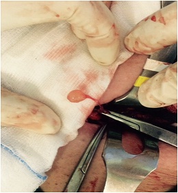

Figure 1: Intraoperative photograph showing the cyst
Peritoneum was opened and hernia defect was identified ((Figure 2) . Round ligament was identified along with the hydrocele of the canal of Nuck. Cyst of canal of nuck was separated from the round ligament and excision of the cyst of canal of Nuck was carried out (Figure 1) . A plug and a 15 × 10 cm polypropylene mesh was placed to cover the hernia defect . Peritoneal flaps were approximated. Post-operative period was uneventful and patient recovered satisfactorily. Histopathologic examination confirmed it as Hydrocoele of canal of Nuck. Patient is asymptomatic on follow up.

Figure 2: Intraoperative photograph showing the hernia sac and defect
Discussion
Hydrocele of the canal of Nuck is a rare entity in clinical practice and is an unusual diagnosis with only about 400 reported cases [4]. It is commonly mistaken for inguinal hernia as one-third of the cases of the former are concomitantly present with the latter.
The canal of Nuck was first described by the Dutch anatomist Anton Nuck in 1691. The processus vaginalis within the inguinal canal in women is, therefore, called the canal of Nuck. The Hydrocele of the canal of Nuck is a very rare condition and results from the failure of obliteration of the distal portion of the canal which forms a fluid containing sac. The Hydrocele of the canal of Nuck generally manifests as a painless swelling in the inguinal area and labium in adolescent age groups. Occasionally, it may present with dull aching pain. The size of the lesion is usually small, averaging about 3 cm in length and about 0.3-0.5 cm in diameter [5].
Ultrasound is the preferred investigation, the lesion is typically seen as a well-defined hypoechoic or anechoic mass lying superficially and medial to the pubic bone in the inguinal canal with enhanced posterior through translucency. It may show cystic in appearance as in our case, or with septations within the lesion [6].
Magnetic Resonance Imaging can give more precise images including septation and a communication between cystic lesion and peritoneal cavity and information on the anatomical relation with adjacent structures. Therefore, MRI can help to diagnose in patients with inguinal cystic mass [7].
The differential diagnosis of a cystic mass in the female groin region includes round ligament cysts, varicosities of the round ligament, inguinal herniation of the ovary, cystic lymphangiomas, epidermal inclusion cysts, abscesses and pseudoaneurysms [8]
The final diagnosis is made during surgery and confirmed by pathological examination. Surgical resection of the hydrocele and ligation of the neck of the processus vaginalis should be considered as standard therapy [1].
Open excision of the cyst of canal of Nuck has been the standard of care, however laparoscopic excision has been reported in the literature.
Authors contribution
VL: Concept and Design, data collection, analysis and interpretation of data, drafting, revision and final approval of the article.
DGC: Concept and Design, data collection, analysis and interpretation of data, drafting, revision and final approval of the article.
GP: Concept and Design, data collection, analysis and interpretation of data, drafting, revision and final approval of the article.
MT: Concept and Design, data collection, analysis and interpretation of data, drafting, revision and final approval of the article.
GC: Critical revision and final approval of the article.
FI: Critical revision and final approval of the article.
Competing interests
The authors declare that there are no competing interests
Ethical considerations
The written informed consent was obtained from the patient and the copy of consent is available with the authors.
Funding:
None
References
[1)Choi YM, Lee GM, Yi JB, Yoon KL, Shim KS, Bae CW, et al. Two cases of female hydrocele of the canal of nuck. Korean J Pediatr. 2012;55:143–6.[Pubmed]
[PMC Full Text]
[2]Park SJ, Lee HK, Hong HS, Kim HC, Kim DH, Park JS, et al. Hydrocele of the canal of Nuck in a girl: Ultrasound and MR appearance. Br J Radiol. 2004;77:243–4.[PubMed]
[3]Qureshi NJ, Lakshman K. Laparoscopic
excision of cyst of canal of Nuck. J Minim Access Surg. 2014 Apr;10(2):87-9.
doi: 10.4103/0972-9941.129960. [PubMed]
[PMC Full text]
[4]Jagdale R, Agrawal S, Chhabra S, Jewan SY. Hydrocele of the canal of Nuck: Value of radiological diagnosis. J Radiol Case Rep. 2012;6:18–22.[PubMed]
[PMC Full text]
[5Park S.J., Lee H.K., Hong H.S. Hydrocele of the canal of Nuck in a girl: ultrasound and MR appearance. Br J Radiol. 2004;77:243–244 [PubMed]
[6]Walter H.S., Martin M. Female hydrocele (cyst of the canal of Nuck) J Ultrasound Med.2004;23:429–432 [PubMed]
[7]Manjunatha YC, Beeregowda YC, Bhaskaran A. Hydrocele of the canal of Nuck: Imaging findings. Acta Radiol Short Rep. 2012;1:12–5 [PubMed]
[PMC Full text]
[8]Caviezel A, Montet X, Schwartz J, Egger
JF, Iselin CE. Female hydrocele: the cyst of Nuck. Urol Int. 2009;82(2):242-5.
doi: 10.1159/000200808. Epub 2009 Mar 19.[PubMed]

