Original Article

Subpial spinal lipoma without dysraphism
*Vivek Sharma (M.Ch.),* *Divye Prakash Tiwari(M.Ch.)* *Janak Raj(M.S.)*
- **Department of Neurosurgery, Institute of Medical Sciences,Banaras Hindu University, Varanasi-221005,India
- Friday, November 01, 2013
- Thursday, November 14, 2013
- Wednesday, November 20, 2013
This is an Open Access article distributed under the terms of the Creative Commons Attribution License (http://creativecommons.org/licenses/by/3.0), which permits unrestricted use, distribution, and reproduction in any medium, provided the original work is properly cited.
Abstract
Introduction: Subpial spinal lipoma without dysraphism is rare lesion and the management remains challanging. In our study we present the extensive clinico-radiological findings and optimum management protocol.
Methods
Fifteen patients who reported with nondysraphic spinal cord lipomas between March 2000 and April 2012 were retrospectively reviewed. All had varying degrees of neurological symptoms at the time of surgery with characteristic features on magnetic resonance imaging. All patients underwent decompression with a laminectomy/laminoplasty and duroplasty.
Results
The age at presentation ranged from 14 to 45 years (mean 28.8Yrs). Patients were followed-up for minimum of 6 months and maximum of 5 years. The most common location was dorsal spine while 4 vertebral segments involvement was found in 66.6% of cases. There was neurological improvement following surgery in all cases. Four patients had recurrence of symptoms.
Conclusion
Surgical excision of subpial lipomas is best treatment offered to the patients presenting with neurological deficits. The amount of surgical resection has not any influence on recovery from symptoms. Hence aggressive debulking should not be attempted as it may aggrevate neurological deficit. Thus adequate decompression with preservation of neural structures should be the aim of surgery.
Keywords
Intraspinal subdural lipomas, Intradural lipoma, Non-dysraphic lipoma, Spinal lipoma,
Introduction
Spinal cord lipomas not associated with spinal dysraphism are rare and make up less than 1% of all spinal cord tumors [1.Lipoma are benign tumor and histologically identical to normal body fat. Lipomas of the spinal cord are frequently associated with spina bifida (lipomyelomeningocoele). These are more commonly located in the lumbosacral or cervical region, and it is due to closure of terminal site of embryonic neural arch, with abnormal inclusion of adipose tissue within the closing lips of the neural groove. Intraspinal lipoma can be broadly divided into Intradural tumor which is intimately associated with the structure of the spinal cord and Epidural lipoma[2].Gowers published the first report of an intradural lipoma in cases of intradural lipoma extending the entire distance of the spinal canal [1]but the most frequent site is the conus medullaris as a component of the dysraphic state.Intraspinal subdural lipomas with spinal dysraphism are commonly found in childhood, mostly located in lumbosacral region. In contrast, the subpial lipomas without dysraphism believed to be congenital in origin, mainly located at cervicodorsal segment. It presents with features of intradural lesion and could be found at all levels of spinal cord with wide age distribution [4].
Patients and Methods
All patients of subdural spinal lipoma among all spinal lesions were taken into consideration. The clinical, radiological and treatment analysis was done. The optimum technique of decompression was reviewed. The outcome with reference to partial versus aggressive decompression is discussed in interest of quality of life of patient.
Results
Since March 2000 and April 2012, we have encountered fifteen cases of subpial spinal cord lipomas without evidence of dysraphism. The female versus male ratio was 9:6. The age at presentation ranged from 14 to 45years (mean 28.8). Minimum follow-up was 6 months and maximum follow-up was 5 years. [Table 1]
| Age Group |
Sex |
Number of Cases |
Percentage |
| 10-20 |
Male
Female
|
1
1
|
6.6
6.6
|
| 20-30 |
Male
Female
|
4
6
|
27
40
|
| 30-40 |
Male
Female
|
1
1
|
6.6
6.6
|
| 40-50 |
Male
Female
|
0
1
|
0
6.6
|
Four patients in our series presented with a short history of worsening neurological symptoms. Six patient had bowel and bladder symptoms. Two patients reported to our institution very late due late referral with grade o power. The quadriparesis was found in 4 while paraplegia. There was grade seven patients in grade 4 power. [Table 2]
| Name of clinical feature |
Number of patient |
Percentage |
| Pain in back |
15 |
100 |
| Spinal deformality |
7 |
46.6 |
| Paraplegia |
11 |
73.3 |
| Quadriparesis |
4 |
26.6 |
| Bladder and System |
6 |
40 |
All patients had pre-operative magnetic resonance imaging (MRI) of T1, T2 and fat separation sequences [Fig 1,Fig2,Fig3] and were diagnosed as case of spinal cord lipoma. In two cases location of tumour was in C2-C5 while in other two patients, cervico-thoracic was the site lesion. Lesion was entirely thoracic in 11 cases. The segment of vertebral body involvement ranged from 3 to 6 with 4 in nine cases. [Table 3].
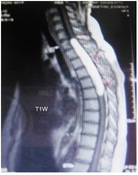
Figure 1: Sagittal MRI T1 weighted
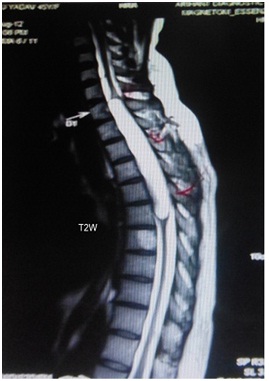
Figure 2: Sagittal MRI T2 weighted
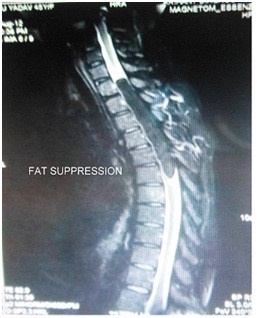
Figure 3: Sagittal MRI fat separation
| Spread of lesion in reference to vertebral body |
Number of cases |
| 6 |
4(26%) |
| 4 |
9(60%) |
| 3 |
2(13.3%) |
The aim of surgery was maximum decompression preserving the neurological function. Four patients subjected to laminectomy and eleven had laminoplasty. In all cases, the lesions presented to the surface and were covered by a thin layer of pia (subpial). The CUSA was used for debulking and standard microneurosurgical principles were used during surgery.The surgical strategy was not to attempt to develop any interface between the spinal cord and the tumor as there was no clear plane identifiable between the lipoma and cord [Fig 4] performed using tensor facia lata as dural graft. There was no documented post operative complication in cases where subtotal decompression was done. The aggressive decompression was tried in 3 cases but with poor outcome. Histopathological report were lipoma .All except 3 patients had improvement in symptoms and neurological status following surgery in the regular follow up period. [Table 4]
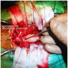
Figure 4: Per-operative View of Subpial Lipoma
| Degree of decompression |
Number of cases |
Postoperative outcome |
| Near total excision |
3 |
Bad |
| Subtotal excision |
8 |
Good |
| Partial excision |
4 |
Partial improvement |
Discussion
Subpial lipomas of spinal cord are most common dysembrogenic lesion accounts for 1 % of intramedullary tumors with embryogenic basis [5].Some authors have postulated that these lesions are due to inclusion of embryogenic rests of fat cell during neural tube formation whereas second school of thought for etiology is connective tissue metaplasia due to premature dysjunction of neural tube from superficial ectoderm [6].It is therefore not a true neoplasm but a hamartoma or a malformation. This can explain why the lipoma is dorsally located and may also explain spinal lipoma without dysraphism .There can be peripheral nerve twig, dermoid cyst, skeletal muscles and lymphoid or renal tissue within the fat. These originate from ectoderm or mesoderm [7].Although all of these theories share some basic aspect, they do not fully explain the exact genesis of the spinal lipoma. Spinal cord lipomas seem to have a peak presentation between 10 and 40 years, with a slight male preponderance. The usual presentation is of a long indolent history, followed by a dramatic worsening over months. Non-dysraphic intradural lipomas are commonly found in the thoracic spine, followed by the cervico-dorsal region with only 12% in the cervical cord alone [8].he characteristic location is in the posterior aspect of the cord within a small radius around the midline. They tend to be present to the surface (subpial), and distort and expand the spinal cord (juxtamedullary). Most of them involve several spinal cord segments. True intramedullary lipoma is very rare with few case series reported in the literature [9 Fifty-five percent of all cases of intradural lipomas present in their second or third decades of life. Around 24% of patients present in their first decade and 16% during the fifth decade. They are equally prevalent in both sexes. The presentation is often with an ascending spastic paresis affecting one or both legs. Pain when present is usually not radicular but rather localised to the area involved. Most patients are symptomatic for 2 years before seeking medical advice. However, in cases of lipomas restricted solely to the cervical spinal cord, over 80% of patients displayed symptoms for more than 10 years [10].During the first decade of life, spinal lipomas may present as tetraplegia or floppy baby syndrome; the neurological manifestations are thought to be due to birth trauma to the spinal cord. In other patients, however, the manifestation is often delayed [11].Almost always these lipomas are located at posterior aspect of spinal cord. The diagnostic modality of choice is MRI due to high sensitivity and because of the high proportion of fat, there is a short T1 relaxation time. Thus, lipomas have characteristic hyperintense appearance on T1-weighted images.This can be confirmed with fat suppression images. Benign lipomas have relaxation parameters similar to those of subcutaneous fat. Liposarcomas and other fat-related tumors have longer T1 relaxation times and therefore are less intense than subcutaneous fat on T1-weighted images. Subpial lipomas are adherent to the cord and nerve roots tightly and difficult to dissect and hence the most effective treatment in still unclear. Some have suggested that since the fat of the lipoma is metabolically identical with normal body and the patient should undergo aggressive weight loss and scrupulous control of diet [12]. This clinical approach to control the growth of the intradural lipoma is controversial as many have observed rapid growth despite strict weight control. Endoh et al. reported a case of an intradural lipoma in a 9-year-old boy that shrank spontaneously with loss of general body fat [13].The aim of surgery should be decompression with preservation of neurological function. The percentage of tumor debulked does not necessarily correlate with the clinical outcome. Good outcome has been reported with laminectomy and biopsy alone [14]. The overwhelming neurosurgical opinion regarding treatment of spinal cord lipoma is that surgical debulking of the tumour is the goal, and not excision. Indeed, total excision has generally shown less post-operative improvement, and in some cases significant worsening of symptoms [15]. Following review of the literature and from our own experience, we propose that asymptomatic patients, and patients with local symptoms only, e.g. pain with no neurological signs & symptoms can be managed conservatively with regular clinical and radiological monitoring. We feel that subtotal removal of the tumor using microscope and CUSA with dural patch grafting to enlarge the space for the slow growing residual tumor is a prudent and safe approach. Similar approach is adopted by the other author as well.
Conclusion
Subpial lipomas without dysraphism are very uncommon commonly affecting dorsal region presenting as slowly progressive neurological deficit It is adherent to the cord and nerve roots hence subtotal removal of tumor using microscope, CUSA or CO2 laser with duroplasty is the treatment of choice. Surgical aspirator is absolutely essential as tumor is not suckable. Instability of spinal column in case of long segment laminectomy is avoided by double door laminoplasty technique.
Authors' Contribution
VS: Concept and design, editing of the final manuscript
DPT: Literature review and preparation of manuscript.
JR: Data collection and preparation of the manuscript.
Conflict of Interests
The authors declare that there are no conflicts of interests.
Ethical Considerations
This is a retrospective review and hence exempted from ethical committee review.
Funding
None declared
Acknowledgement
None
References
[1].Caram PC, Scarcella G, Carton CA. Intradural lipomas of the spinalcord. With particular emphasis on the intramedullary lipomas. J Neurosurg 1957; 14:28–42.[pubmed]
[2].Liebeskind et al. Intraspinal lipomas. Neuroradiology 1974; 7(4): 198–200[pubmed]
[3].Gowers WR. Myo-lipoma of the spinal cord. Trans Soc Path Soc Lond 1897; 27: 19-22.
[4].Harjinde Singh Bhatoe, M. Ch., Prakash Singh, M. Ch. Aarti Chaturvedi et al, Nondysraphic Intramedullary Spinal cord Lipoma: A Review, Neurosurg Focus 2005; 18 (2)[pubmed]
[5].Schwartz TH, McCormick PC: Intramedullary tumors of the spinal cord, in Batjar HH, Loftus CM (eds): Textbook of Neurological Surgery: Principles and Practice, Philadelphia: Lippincott Williams& Wilkins, 2003, Vol 2, pp 1864-1871.
[6].Huda MF, Mohanty S, Sharma V et al Subpial lipoma of the spinal cord. Indian J Radiol imaging 2003; 13:423-6.
[7].Ammerman BJ, Henry JM, De Girolami U, Earle KM. Intradural lipomas of the spinal cord. A clinicopathological correlation. J Neurosurg 1976, 44:331–336[pubmed]
[8].Lantos G, Epstein F, Kory LA. Magnetic resonance imaging of intradural spinal lipoma. Neurosurgery 1987, 20:469–472.[pubmed]
[9].Syed Mohammed Rezaul Kabir &Dominic Thompson&Kia Rezajooi &Adrian T. H. Casey. Non-dysraphic intradural spinal cord lipoma: case series, literature review and guidelines for management. Acta Neurochir 2010, 152:1139–1144.[pubmed]
[10].Giuffre R. Intradural spinal lipomas. Review of the literature (99 cases) and report of an additional case. Acta Neurochir (Wien) 1966, 14:69–95.[pubmed]
[11].Bhatoe HS, Singh P, Chaturvedi A, Sahai K, Dutta V, Sahoo PK . Nondysraphic intramedullary spinal cord lipomas: a review. Neurosurg Focus, 2005;18:ECP.[pubmed]
[12].Van Dellen JR, Van Den Heever CM. Intraspinal Lipoma. A case report S Afr Med J 1976; 50-49-50. [pubmed]
[13]Endoh M, Iwasaki Y, Koyanagi I, Hida K, Abe H. Spontaneous shrinkage of lumbosacral lipoma in conjunction with a general decrease in body fat: case report. Neurosurgery 1998; 43:150–151.[pubmed]
[14].Ammerman BJ, Henry JM, De Girolami U, Earle KM. Intradural lipomas of the spinal cord. A clinicopathological correlation. J Neurosurg 1976; 44:331–336.[pubmed]
[15].R. Pathi, M. Kiley,.M. Sage. Isolated spinal cord lipoma Case report. Journal of Clinical Neuroscience 2003;10(6), 692–694[pubmed]


