Original Article

Transperitoneal laparoscopic management of ureteric stones: A prospective study
* Muneer Ahmed Para , *Reyaz Ahmed Wani
- * Postgraduate Department of Surgery, Government Medical College Jammu, Jammu and Kashmir, India
- Submitted: September 10, 2020
- Accepted: November 11, 2020
- Published:: November 11, 2020
This is an Open Access article distributed under the terms of the Creative Commons Attribution License (http://creativecommons.org/licenses/by/4.0), which permits unrestricted use, distribution, and reproduction in any medium, provided the original work is properly cited
Abstract
Background
Ureteral calculi affect a large section of population and most of them are symptomatic. Depending on the size and location of stone, the treatment can range from observation, pharmacotherapy, endourological intervention, shock wave lithotripsy (SWL), laparoscopic or open retrieval. Though the indications of open and laparoscopic interventions are declining with advances in endourology, these options are still considered in large impacted stones. We share our experience with laparoscopic transperitoneal ureterolithotomy for large and difficult ureteric stones.
Methods
A prospective study over a period of one year was undertaken at a tertiary care government centre in North India. The patients of ureteric calculi were enrolled on both emergency and outpatient basis. After evaluation, a set of patients (Age >> 17 years; Stone size >15mm) were selected for laparoscopic transperitoneal ureterolithotomy. The outcome was recorded in terms of operative time, complications, stone clearance rate and hospital stay.
Results
A total of 30 patients underwent laparoscopic transperitoneal ureterolithtomy. The main complaints were abdominal pain in about 80%, vomiting in 60%, dysuria in 11% and hematuria in 10% patients. The stones ranged in size from 16 mm to 25 mm with average size of 19.5 mm. All patients were operated by single surgeon with a mean operative time of 82.60 minutes. There were no major perioperative complications with a stone clearance rate of 100%. Two patients had minor complications in the form of postoperative fever and paralytic ileus, which were managed conservatively. Most of the patients were discharged on or before 3rd postoperative day.
Conclusions
Laparoscopic transperitoneal ureterolithotomy is a good treatment modality for large stones not feasible for ureteroscopic removal or SWL. Besides the advantages over open surgery, MIS has very high stone clearance rate and minimal complications.
Keywords
Ureteral calculi, ureteroscopy, Shock wave lithotripsy (SWL), intravenous urogram (IVU), stone clearance rate
Introduction
Renal stone disease is one of the most common afflictions of modern society. The lifetime prevalence of kidney stone disease in US is 1-15%, with the probability of having a stone varying according to age, gender, race and geographic location. In India, approx. 57 million people suffer from stone disease and at least 1/1000 of population needs hospitalization due to kidney stone disease every year. Various risk factors for stone formation have been studied; most common ones include family history with polygenic inheritance, dietary and environmental factors. The diagnosis of upper tract calculus is not difficult in most patients. All patients in who stones form should be evaluated to detect treatable causes and to prevent recurrences.
Although the traditional indications for intervention (intractable symptoms, infection, obstruction and a stone that is unlikely to pass spontaneously) have not changed, the advances in minimal invasive surgery allow almost any symptomatic patient to be considered a candidate for stone removal. A thorough knowledge of the natural history of ureteral calculi is important to decide about the management (conservative versus intervention). In the absence of external ureteral compression or internal narrowing, the width of the stone is the most significant measurement affecting the likelihood of stone passage. In stones smaller than 4mm, 4-6 mm and larger than 6mm, the rates of spontaneous passage of 80%, 59% and 21%, respectively have been seen [1]. However, the measurement of stone size from a plain radiograph can be misleading. Otnes and Sandnes (1978) reported that the stone size was overestimated in 59% of cases, underestimated in 15% and correlated accurately with the actual size in only 26% [2]. Rate of spontaneous passage also depends on stone location; passage rates from the proximal, middle and distal ureteral stones have been found to be 22%, 46% and 71%, respectively [3]. Segura and associates (1997) reported in the American Urological Association (AUA) guidelines on the management of patients with ureteral calculi that for stones smaller than 5mm, the spontaneous passage rate in the distal ureter and proximal ureter ranged from 71-98% and 29-98%, respectively; whereas stones larger than 5mm had a lower spontaneous passage rate ranging from 10-53% and 25-53% for proximal and distal ureteral calculi, respectively [4]. Therefore, for patients with stones of 5 mm or less, conservative management should be considered, whereas the chance of spontaneous passage for larger stones diminishes considerably, and intervention should be more readily contemplated. Besides size and site of stones, duration of symptoms also helps to predict the need for surgical intervention. Several studies have found that if significant progress has not occurred after one month of observation, intervention is usually required [5]. Furthermore, ureteric stone of any size may be associated with renal obstruction, and care must be taken to prevent irreversible damage to the kidney, whether the patient selects expectant or active treatment.
The treatment of urinary lithiasis has been revolutionized during the last three decades. Minimally-invasive therapies in the form of endoscopic surgery in conjunction with shock wave lithotripsy (SWL) have diminished the role of open stone surgery. Advantages of laparoscopic method over open method include cosmesis, lesser post-operative pain, lesser wound complications, blood loss and the length of hospital stay. Thus laparoscopy is continuously gaining place in the treatment of urinary stones, mainly replacing open surgery [6]. Laparoscopic ureterolithotomy is mostly recommended for large impacted stones or when endoscopic ureterolithotripsy or SWL have failed (Campbell-Walsh Urology 10th Edition). Both retroperitoneal and transperitoneal approaches for laparoscopic ureterolithotomy have been described. We present our initial experience with laparoscopic transperitoneal ureterolithotomy.
Patients and Methods
This prospective study was conducted in Postgraduate Department of Surgery, GMC Jammu from November 2012 till October 2013. All patients with age >14 years, having large (>1.5 cm) ureteric stones from ureteropelvic junction to upper border of sacroiliac joint who presented in emergency or on outpatient basis were included in the study. After thorough history and physical examination, patients were subjected to following investigations: Urine routine examination, complete blood count, coagulation profile, kidney function test, ultrasonography (USG), intravenous urogram (IVU) and DTPA scan (optional). A plain X ray abdomen (KUB) was taken in the morning on the day of surgery to confirm the exact site of stone. Transperitoneal laparoscopic ureterolithotomy was accomplished through 3 ports, a 10mm camera port placed 2 fingerbreadths lateral and superior to umbilicus on the contralateral side, 10 mm and 5 mm working ports placed a handbreadth superior and inferior to camera port, respectively. A vertical ureterotomy over the stone was followed by stone extraction in glove finger via 10mm working port to prevent spill. The ureterotomy was closed with vicryl 4-0 suture over a DJ stent. A tube drain was placed via 5mm port if deemed necessary. Postoperative X ray KUB was taken in the morning of 1st postoperative day to look for stone clearance and stent position. Stone clearance was also confirmed with the help of USG in doubtful cases. The patients were discharged once ambulatory and on full oral diet and medication. The stent was removed after 4-6 weeks’ time.
Results
A total of 30 patients underwent laparoscopic transperitoneal ureterolithotomy, which included 26 males and 4 females. Majority (2/3) of patients were between 20-40 years age. Average age of presentation was 31 years. Youngest patient was 21 years old and oldest one 55 years old. Stones were located at L3-L4 in 22 (73.3%) and at L4-L5 in 8 (26.7%) patients (Figure 1). In our study, 17 patients (56.67%) had stones on left side while 13 (43.33%) had on the right side. Colicky abdominal pain was the predominant symptom seen in 24 (80%) patients. Other symptoms were vomiting in 18 (60%), burning micturition in 11 (36.7%) and hematuria in 10 (33.3%) patients. Duration of symptoms ranged from 6 hours to more than one year. 60% patients presented within 2 months after onset of symptoms. Average size of calculi in our study was 19.5mm, largest measuring 25mm and smallest of 16mm. Average operative time was 82.60 minutes. Postoperative analgesic requirement was 2-3 doses of 75 mg injectable diclofenac on day one and thereafter 1-2 days of oral diclofenac. In the immediate post-operative period, 63% patients needed 150 mg injectable diclofenac (2 doses of 75 mg each); while 37% patients needed 225 mg (3 doses). All the patients were stone free after the procedure as confirmed by X ray KUB. There were no major complications; while two Minor complications reported were fever in one patient and paralytic ileus in another. More than 85% patients were discharged on 3rd postoperative day. Average hospital stay was 4 days with a maximum of 6 days and minimum of 3 days.
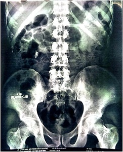

Fig 1. X ray KUB showing ureteric calculus
Discussion
Since the introduction of SWL and ureteroscopy for the management of ureteric calculi, the routine use of an open surgical approach for removal of ureteric calculi has rapidly declined. However, large ureteric calculi pose significant challenge for modern endourologic techniques, often requiring several endoscopic procedures as well as SWL sessions. SWL is found to be suitable for managing ureteric stones of <1cm. As the stone size increases, the chance of clearance decreases to 60% of the need for multiple sessions increases [7]. Park et al (1998) [7] reported that the stone free rate decreased from 84% to 42% when the stone is >1 cm. Thus, the indications for laparoscopic ureterolithotomy in the age of modern endourology include stones which cannot be assessed ureteroscopically or cannot be fragmented by SWL.
Laparoscopic ureterolithotomy is a minimally invasive option to treat large ureteric stones not amenable to ureteroscopy (Figure 2 3 4 5 -6). In our study, transperitoneal approach has been described for stones from PUJ to upper border of sacrum. It gives better understanding of the anatomical landmarks particularly for the ureteric stone. The first transperitoneal laparoscopic ureterolithotomy was performed by Raboyet al in 1992 [8]. Gaur (1992) described retroperitoneal laparoscopic approach for ureteral calculi facilitated by hydraulic balloon dilatation system [9]. Thereafter, several authors have tried to replace open ureterolithotomywith either a transperitoneal or a retroperitoneal laparoscopic procedure [10-13].

Fig 2. Ureterotomy by monopolar hook

Fig 3. Extraction of stone from ureter

Fig 4. Placement of stone in glove finger

Fig 5. Placement of DJ stent

Fig 6. X ray KUB showing stent in place with no residual stone
The commonest age reported by most of the workers (Higgins 1939, Bumpus and Thomson 1925) for presentation of ureteric calculi is between 20 and 50 years [14, 15]. It is rare in childhood and unusual in old age [16]. In our study, the average age of presentation is 31 years. Most of the studies have reported a male to female ratio of 3:1 and 2:1. Cleveland clinic reported a ratio of 80% males to 20% females. In our study, there were 86.66% males and 13.33% females. Pain, nausea/vomiting, burning micturition, haematuria were the most common symptoms in the present study. Higgins (1939) [14] reported 50% incidence of colic; whereas in a study by Miles et al (1998) 92% patients had colic and 22% had haematuria [17]. In our study, 80% patients had colicky pain, 36.6% had burning micturition. This is almost similar to other studies. Miles Fox et al (1965) [18] reported that duration of symptoms varies from 3 hours to 5 years. 80% patients came within one month of onset of symptoms, while 4% had a history of one year or longer. In this series, 60% of patients came in two months after appearance of symptoms and only 40% reported between 3 months and one year. SWL is found to be suitable for managing ureteric stones of <1 cm. As the stone size increases, the chance of clearance decreases and the need for multiple sessions increases. Park et al [7] reported that the stone free rate decreased from 84% to 42% when the stone size is > 1cm. Thus, the indications of laparoscopic ureterolithotomy in the age of modern endourology include stones which cannot be accessed ureteroscopically or cannot be fragmented. So, we selected the patients with calculi more than 15 mm. Average size of calculi in our study is 19.5 mm. Largest measuring 25 mm and smallest of 16 mm. Bumpus and Thompson (1925) [15]found calculi with equal frequency on either side. In the present series, 43.33% were on the left side while 56.67% on the right side. Duration of procedure depends on many factors like whether the stone is impacted, recurrent, previous procedures and most importantly expertise of the surgeon. Opening the ureter (ureterotomy) is the main controversial issue. Nouriaet al., [19] have recommended that use of a cold knife for opening the ureter is a wiser choice to prevent stricture. Muslumanogluet al., [20], however, suggested that an electric hook using cutting current was much easier. The use of diathermy hook electrode to open the ureter was reported as a safe method by Harewood et al., [10]. Other technical considerations include providing an immobile ureter during the procedure, bleeding from the ureteral wall and severe adhesions around the stone can make the procedure difficult. One more thing which takes more time and requires experience is intracorporeal knotting. Many studies showed that the operative time ranging from 85 minutes to 145 minutes. In a study by Abolyosr (2007), the mean operating time was 85.2 minutes,similarly in another study by Skrepetis K, Doumas K (2001), the mean operating time was 130 minutes [12,21]. The mean operating time in some other studies was 145+/- 42 minutes (El-Feel A et al 2007),105 minutes(Keeley et al 1999) and 121.38 minutes (Kijvikai K et al 2006) [22-24]. In our study, the average duration of surgery was 82.60 min. with a maximum of 118 minutes and a minimum of 48 minutes. Duration of procedure was more in initial patients and less in those patients which were operated at the end of our study. This was probably due to the learning curve effect. Postoperatively, we treated the pain with intramuscular injection of Diclofenac. Majority of patients received two doses of diclofenac, 75 mg each dose. This was followed by oral tablet diclofenac 50mg once the patients started taking liquids orally. Pain severity was assessed according to visual analogue score (VAS). All of the patients received analgesics on first postoperative day - 63% received 150 mg (75 mg in 2 doses) and 37% required 225 mg (75 mg in 3 doses). When compared to open procedure, analgesic requirement in laparoscopy is significantly less. In Skrepetis K, Doumas K (2001) [12] study, analgesic medication requirement per patient was one day for transperitoneal laparoscopic ureterolithotomy (TLU) and 4 days for open ureterolithotomy (OU). In El-Feel a et al (2007) [22] study, postoperative analgesia was single dose of a non-steroidal anti-inflammatory on day one. In our study, all the patients were started orally 6 hour postoperatively. All patients made uneventful recovery except for two patients. One developed postoperative ileus and other fever. The patient with ileus was kept NPO and started on IV fluids and antibiotics and the patient stayed for longer duration compared to others (6 days). Other patient responded to IV antibiotics and stayed in hospital for 5 days. Analgesics were given for 2 days; on 1st postoperative day intramuscular diclofenac and on 2nd postoperative day oral diclofenac. Urinary catheter was removed on 2nd day and patients were observed till they passed urine. Most of the patients were discharged on the 3rd day. No patient needed blood transfusion postoperatively. Other studies showed that the mean hospital stay was 3.8 days [21], 3 days [12], and 4.1 +/- 6.7 days [22]. Matias DB et al [25] showed the mean overall hospital stay was 3.3 days. Similarly, the postoperative hospital stay ranged from 2-5 days (mean 3 days) by Flasko T et al.,[26]. In our study, average postoperative hospital stay of the patients is 4 days, minimum of 3 days and maximum of 6 days. Perioperative complications in our study were seen in only 2 patients - one who developed postoperative ileus which we managed conservatively. Other developed fever which responded to antibiotics. There were no SSI, residual stone, stent displacement or other stent related complications or stricture formation on follow up. In the study by Abolyosr A, no major intra or postoperative complications except prolonged urine leak in 2 patients, which lasted for 7 and 9 days and resolved with conservative management only[21]. One more study done by Skrepetis K, Doumas K, postoperative complications included urinary leak, subcutaneous haematoma, subcutaneous emphysema in the TLU group and wound site and urinary tract infections in the OU group [12]. Study by Simforoosh N et al., (2007) showed that on the first postoperative day, 119 (96.7%) patients were stone free [27]. Surgical complications occurred in 14 (11.4%) patients and conversion to open surgery was required in one (0.8%) due to migration of the calculus to the peritoneum after removal from the ureter. Re-operation was carried out in 1 patient (0.8%). There were 3 cases (2.4%) of urinoma, all responded to DJ stent insertion.
Conclusion
The surgical treatment of ureteric calculi is governed by stone characteristics (size, location), patient characteristics (symptom severity, duration and associated morbidity) and technical expertise available. Despite availability of SWL and advances in endoscopic management of ureteral stones, large impacted stones still need alternative treatment modalities to avoid the risk of multiple procedures/sessions and high cost. Open ureterolithotomy is still a common practice in developing countries; however, Laparoscopic ureterolithotomyis taking over.
Author’s contribution
MAP: Conceptualization, study design, data
collection and analysis
RAW: Study design, data collection and
drafting for publication
Ethical Considerations and consent
The study is approved by Institutional ethical committee and written informed consent was obtained from all cases. The copy of ethical approval and consent is available with authors.
Conflict of Interests
The authors declare that there are no conflicts of interests
Funding
None
Acknowledgement
None
References
[1].Ueno A, Kawamura T, Ogawa A, et al. Relation of spontaneous passage of ureteral calculi to size. Urology 1977;10:544-46[PubMed]
[2]Otnes B, Sandnes H. Comparison of radiological measurement and actual size of ureteral calculi. Scandinavian Journal of Urology and Nephrology 1978;12(2):155-56[PubMed]
[3]Morse RM, Resnick MI. Ureteral calculi: natural history and treatment in an era of advanced technology. J Urol1991;145:263-65[PubMed]
[4]Segura JW, Preminger GM, Assimos DG, et al. Ureteral stones clinical guidelines panel summary report on the management of ureteral calculi. The American Urological Association. J Urol1997;158:1915-21[PubMed]
[5].Ibrahim AI, Shetty SD, Awad RM, et al. Prognostic factors in the conservative treatment of ureteric stones. Br J Urol1991;67:358-61[PubMed]
[6].Hruza M, Schulze M, Teber D. Laparoscopic techniques for removal of renal and ureteral calculi. J Endourol 2009;23(10):1713-18[[PubMed]
[7.Park H, Park M, Park T. Two-year experience with ureteral stones: Extracorporeal shock wave lithotripsy versus ureteroscopic manipulation. J Endourol1998;12:501-4[PubMed]
[8]Raboy A, Ferzli GS, Ioffreda R, Alber PS. Laparoscopic ureterolithotomy. Urology 1992;39(3):223-25[PubMed]
[9].Gaur D D. Laparoscopic operative retroperitoneoscopy:use of a new device. J urol 1992;148(4):1137-9[PubMed]
[10].Harewood LM, Webb Dr, Pope AJ. Laparoscopic ureterolithotomy: the results of an initial series, and an evaluation of its role in the management of ureteric calculi. Br J Urol 1994;74(2):170-6[PubMed]
[11].Simforoosh N, Basiri A, Danesh AK. Laparoscopic management of ureteral calculi: a report of 123 cases. Urol J. 2007;4(3):138-41[PubMed]
[12].Strepetis K, Doumas K, Siafakas I, Lykourinas M. Laparoscopic versus open ureterolithotomy: a comparative study. Eur Urol. 2001;40(1):32-6[PubMed]
[13].Turk I, Deger S, Roigas J, et al. Laparoscopic ureterolithotomy. Tech Urol. 1998;4(1):29-34[PubMed]
[14].Higgins CC. Factors in recurrence of renal calculi. JAMA 1939;113(16):1460-65
[15].Bumpus KC, Thompson CJ. Stones in the ureter. SurgGynecolObstet1925;50:106-9
[16].Drach GW. Urinary lithiasis. Chapter 22pp 779-878 in Campbells urology.m Harrison JH et al (editors) Saunders 1978.
[17].Miles M, Pyrah J. Raper HK. Management of lower ureteric calculi – series of 1061 patients.JEndourol 1998;12(6):501-4
[18].iles F, Pyrah LN, Raper FP. Management of ureteric stone: a review of 292 cases. BJU 1965;37(6):660-70[PubMed]
[19].Nouira Y, Kallel Y, Binous MY, Dahmoul H, Horchani A. Laparoscopic retroperitoneal ureterolithotomy: initial experience and review of literature. J Endourol. 2004;18(6):557-61[PubMed]
[20]Muslumanoglu AY, Karadag MA, Tefekli AH, Altunrende F. When is open ureterolithotomy indicated for the treatment of ureteral stones ? Int J Urol 2006;13(11):1385-88 [PubMed]
[21].Abolyosr A. Laparoscopic transperitoneal ureterolithotomy for recurrent lower ureteral stones previously treated with open ureterolithotomy: initial experience in 11 cases. J Endourol. 2007;21(5):525-9 [PubMed]
[22].El-Feel A, Abouel-Fettouh H, Abdel-Hakim AM. Laparoiscopic transperitoneal ureterolithotomy. J Endourol. 2007;21(1):50-4. [PubMed]
[23].Keeley FX, Gialas I, Pillai M, et al. Laparoscopic ureterolithotomy: the Edinburgh experience. BJU Int. 1999;84(7):765-9 [PubMed]
[24].Kijvikai K, Patcharatrakul S. Laparoscopic ureterolithotomy: its role and some controversial technical considerations. Int J Urol. 2006;13(3):206-10A [PubMed]
[25]Matias DB, Alvim RG, Ribas M. Laparoscopic treatment of ureterolithiasis. ActasUrol Esp. 2009;33(6):667-9 [Pubmed]
[26]Flasko T, Holman E, Kovacs G, et al. Laparoscopic ureterolithotomy: The method of choice in selected cases. J Laparoendosc Adv Surg Tech A. 2005;15(2):149-52 [PubMed]
[27]Simforoosh N, Basiri A, Danesh AK, et al. Laparoscopic management of ureteral calculi- a report of 123 cases. Urology Journal 2007;4(3):138-40 [PubMed]

