Original Article

Laparoscopic and Laparoscopic Assisted Management of Intra- Abdominal Cystic Lesions in Infants and Children.
*Sajad Wani,*, Gowhar Mufti *, Nisar Bhat *, Eajaz Baba
- *Department of Pediatric surgery, SKIMS, Jammu &Kashmir, Srinagar, India
- SubmittedThursday, October 20, 2016
- Accepted:Sunday, January 29, 2017
- Published:Tuesday, January 31, 2017
This is an Open Access article distributed under the terms of the Creative Commons Attribution License (http://creativecommons.org/licenses/by/4.0), which permits unrestricted use, distribution, and reproduction in any medium, provided the original work is properly cited
Abstract
Background
Intra-abdominal cystic lesions in infants and children are relatively common and complete excision of the cyst is the treatment of choice. With the development of laparoscopy, pediatric laparoscopy is being applied to diverse conditions. The aim of this study was to evaluate the laparoscopic management of intraadominal cystic lesions in infants and children using the Tan Bianchi approach.
Material and Methods
Infants and children with different types of intra-abdominal cystic lesions were included in the study. After establishing the pneumoperitonium by open technique, cysts were decompressed and partially or completely dissected depending upon the nature of the cyst. Using the Tan Bianchi approach, cyst was delivered out for final excision or resection.
Results
The age of patients ranged from 4 months to 6 years with mean age of 2.7 years. Fourteen patients were included in the study, which included 6 mesenteric cysts, 3 duplication cysts, 3 omental cysts and 2 ovarian cysts. No intraoperative or postoperative complication was encountered. The mean hospital stay was 3 days.
Conclusion
Laparoscopic or laparoscopic assisted management of intra-abdominal cystic lesions combining withTan Bianchi approach is good alternative to open surgery. It is safe, simple and effective technique even for surgeons with less experience in intracorporeal suturing.
Keywords
Intraabdominal cystic lesions, Laparoscopic, Tan Bianchi umbilicotomy, Infants and children.
Introduction
Intra-abdominal cystic lesions in infants and children are relatively common. Most commonly these arise from the small bowel mesentery, omentum or the ovary. Lymphatic cysts, duplication cysts and parasitic cysts are the common pathological types. Complete excision of these lesions is the treatment of choice and laparotomy has been the conventional approach for resection. The intra- abdominal dissection needed to remove the cyst is limited compared to the trauma of access.
With the improvement in instrumentation and technology, pediatric laparoscopy is being applied to diverse conditions including the management of intra-abdominal cystic lesion in infants and children [1, 2]. It provides an ideal means of decreasing the trauma of access, superior cosmesis, less pain, early recovery, less morbidity and shorter hospital stay. However, in infants and small children with large intra-abdominal cysts, laparoscopy can be challenging due to small intra-abdominal space, large viscera and poor ergonomics in relation to port placement. The aim of this study was to evaluate the safety and efficacy of laparoscopic management of intra-abdominal cystic lesions in infants and children.
Material and Methods
This study was conducted over a period of 3 years from April 2013 to May 2016. Infants and children with different types of intra- abdominal cystic lesions including mesenteric cysts, omental cysts, duplication cysts and ovarian cysts were included in the study and individual cases were managed with laparoscopic assisted surgery using Tan Bianchi approach.
Surgical technique
Under general anesthesia, in all cases, umbilical port was the primary port and was introduced by open technique (Figure 1).
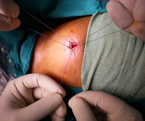
Figure 1 the open method of trocar insertion
Intra-abdominal pressure was maintained at 8 mm Hg in infants and 10-12 mm Hg in older children. Scope was inserted through the 5mm umbilical port. Working ports were placed as required for handling and dissecting the cyst. After confirmation of the diagnosis, cyst was aspirated and dissected either partially or completely depending up on the origin and nature of the cyst. A curvilinear incision was made in the skin at the umbilical port from 3 to 9 O’clock position through 12 O’clock position. Skin was undermined superiorly in the midline. After undermining the skin, a midline cut (3-4 cm) was made in the linea alba (Figure 2).
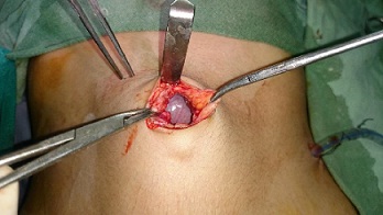
Figure 2 –Excision of cyst
The decompressed cystic lesion was delivered outside the abdomen for excision or resection as required including that of adjacent bowel (Figure 3).
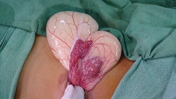
Figure 3 –Excision of adjacent bowel along with cyst
After excision of the cyst, abdominal wall defect was repaired (Figure 4).
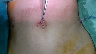
Figure 4 –Repair of abdominal wall post excision cyst
Results
The age of patients ranged from 4 months to 6 years with mean age of 2.7 years. Fourteen patients were included in the study which included 6 mesenteric cysts, 3 duplication cysts, 3 omental cysts and 2 ovarian cysts. Laparoscopic assisted surgery was done in all cases.
There was no intraoperative complication. Excision of cyst was done in all omental cysts, 2 mesenteric cysts and 2 ovarian cysts. Resection with anastomosis was done in all duplication cysts (two colonic and one ileal duplication cyst) and 4 mesenteric cysts. Post-operative period was uneventful and oral intake was started on secondpostoperative day in patients who had undergone resection (Table 1). The mean hospital stay was 3 days and there was no postoperative complication. All the patients are doing well in the follow-up.
Table 1:
| Type of cystic lesion |
Number of patients |
Type of procedure done |
Oral started |
| Mesenteric cyst |
6 |
| Excision in 2 |
| Resection with anastomosis in 4 |
|
|
| Omental cyst |
3 |
Excision in all |
First POD |
| Duplication cyst |
3 |
Resection with anastomosis in all the patients |
Second POD |
| Ovarian cyst |
2 |
Excision |
First POD |
Discussion
Intra-abdominal cysts are categorized based on their location in solid organs, mesentery, omentum, ovary or retroperitoneal. With the increasing use of antenatal ultrasonography, the incidence of neonatal intra-abdominal cysts has increased [3]. The presentation varies from abdominal distension, abdominal pain, palpable abdominal mass or bowel obstruction. They can also present as an acute life threatening intra-abdominal catastrophe such as torsion, bleeding or volvulus.
The optimal surgical management requires complete excision of the cystic lesion. Omental cysts can be excised safely without damage to the adjacent bowel [4, 5]. Mesenteric cysts can be enucleated, however bowel resection is required in 50-60% of cases [6, 7]. If resection or enucleation is not possible because of the size of the cyst or cyst is located deep within the root of the mesentery, partial excision with marsupialization of the remaining cyst can be done. The cyst lining should be sclerosed with electro cautery, 10% glucose solution or tincture of iodine to minimize the recurrence [8]. Partial excision with or without drainage is associated with high recurrence rate [3].
Challenging in infants and children especially in cases of large cyst in small babies, difficulty in access to the peritoneal cavity for port placement, difficulty in dissection and manipulation of the cyst because of limited space and difficulty in ergonomics. We performed the open method of trocar insertion in all patients to prevent the gut or vascular injury as shown in Figure 1.
Chemical peritonitis can occur due to leakage of cyst fluid in to the peritoneal cavity [9]. To prevent this, several laparoscopic approaches and modifications have been described. These include drainage of the cyst under ultrasound guidance, drainage during laparoscopy followed by intracorporeal or extracorporeal excision of the cyst [10-15]. To facilitate the intracorporeal cyst manipulation, we aspirated the cyst via a controlled and direct percutaneous puncture using a spinal needle under laparoscopic view. Using the Tan Bianchi approach (as shown in the Figure 2, cyst was delivered out for excision or resection. Two ovarian cysts were excised intracorporeally which were benign cysts. In our patients there was no intraoperative or postoperatively complication. IIigashimoto et al., reported the advantages of laparoscopic associated surgery in neonates with intra-abdominal cysts [16]. Cohen et al., reported that laparoscopic surgery is suitable for the treatment of adnexal pathology in children [17]. Takayuki et al., reported that laparoscopic associated surgery is effective for the management of omental cysts and ovarian cysts in infants [18].
Laparoscopic associated surgery is safe and effective surgical technique for the management of intra-abdominal cysts in infants and children with good outcome and minimal morbidity. Laparoscopic associated surgery can completely remove the intra- abdominal cystic lesion along with the pedicle and hence preventing the risk of bowel obstruction secondary to remnant of pedicle acting as adhesive band.
Conclusion
With the development of laparoscopic techniques and instrumentation, laparoscopic surgery has become preferred alternative to laparotomy. Laparoscopic associated management of intra-abdominal cystic lesion using Bianchi approach is safe and simple alternative to open surgery with good results and minimal morbidity. By making a midline cut in the linea alba as shown in our technique, intra-abdominal cystic lesions can be brought out easily and hence management becomes much easier. This approach is safe and simple even for surgeons with less experience in intracorporeal suturing.
Authors’ Contribution
SW concieved and designed the study, literature review and mansucript preperation
GM literature review and mansucript preperation
NB literature review and mansucript preperation
EB: design of the study and final editing of the manuscript.
Ethical Considerations
The study was approved by the Institute Ethics committee and individual consent were obtained from all the patients. Copy of consent is available with the authors.
Conflict of Interests
The authors declare that there is no conflict of interests.
Funding
None declared.
References
[1].Waldschmidt J, Schier F. Laparoscopic surgery in neonates and infants. Eur J Pediatr Surg. 1991; 1:145-150s [PubMed]
[2].Delarue A, Guys JM, Louis-Borrione C, Simeoni J, Esposito C. Pediatric endoscopic surgery: pride and prejudice. Eur J Pediatr Surg. 1994; 4: 323-326.[PubMed]
[3]Crombleholme TM, D'Alton M, Cendron M, Alman B, Goldberg MD, Klauber GT, Cohen A, Heilman C, Lewis M, Harris BH. Prenatal diagnosis and the pediatric surgeon: the impact of prenatal consultation on perinatal management. J Pediatr Surg. 1996 Jan;31(1):156-62; discussion 162-3. [PubMed]
[4].Sakurai Y, Taniguchi K, Uyama I, Inaba K, Furuta S, Sunagawa R, Nagasako Y, Ishida Y, Hiramatsu Y, Yonemura J, Isogaki J, Komori Y. Laparoscopic excision of the cystic lymphangioma occurred in the lesser omentum: report of a case and review of literature. Surg Laparosc Endosc Percutan Tech. 2009 Feb;19(1):e11-4. doi: 10.1097/SLE.0b013e31818a8a9b [PubMed]
[5].Nam SH, Kim DY, Kim SC, Kim IK. The surgical experience for retroperitoneal, mesenteric and omental cyst in children. J Korean Surg Soc. 2012 Aug;83(2):102-6. doi: 10.4174/jkss.2012.83.2.102. Epub 2012 Jul 25. [PubMed] [PMC]
[6].de Lagausie P, Bonnard A, Berrebi D, Lepretre O, Statopoulos L, Delarue A, Guys JM. Abdominal lymphangiomas in children: interest of the laparoscopic approach. Surg Endosc. 2007 Jul;21(7):1153-7. Epub 2006 Dec 20. [PubMed]
[7].Vanek VW, Phillips AK. Retroperitoneal, mesenteric, and omental cysts. Arch Surg. 1984 Jul;119(7):838-42 [PubMed]
[8].Antao B, Tan J, Quinn F. Laparoscopic Excision of Large Intra-Abdominal Cysts in Children: Needle Hitch Technique. Case Rep Med. 2015;2015:937191. doi: 10.1155/2015/937191. Epub 2015 Dec 21. [PubMed] [PMC]
[9].Kondo W, Bourdel N, Cotte B, Tran X, Botchorishvili R, Jardon K, Rabischong B, Pouly JL, Mage G, Canis M. Does prevention of intraperitoneal spillage when removing a dermoid cyst prevent granulomatous peritonitis? BJOG. 2010 Jul;117(8):1027-30. doi: 10.1111/j.1471-0528.2010.02580.x. Epub 2010 May 11. PMID: 20465557 DOI: 10.1111/j.1471-0528.2010.02580.x [PubMed]
[10].Morrison CP, Wemyss-Holden SA, Maddern GJ. A novel technique for the laparoscopic resection of mesenteric cysts. Surg Endosc 2002;16: article219 [PubMed]
[11].Schenkman L, Weiner TM, Phillips JD. Evolution of the surgicalmanagement of ovarian cysts: laparoscopic-assisted transumbilical extracorporeal ovarian cystectomy. J Laparoendosc Adv Surg Tech A. 2008 Aug;18(4):635-40. doi: 10.1089/lap.2007.0193.[PubMed] [PMC]
[12].Salem HA. Laparoscopic excision of large ovarian cysts. J Obstet Gynaecol Res. 2002 Dec;28(6):290-4. PMID: 12512924. [PubMed]
[13].Dolan MS, Boulanger SC, Salameh JR. Laparoscopic management of giant ovarian cyst. JSLS. 2006 Apr-Jun;10(2):254-6. [PubMed] [PMC]
[14].Kuga T, Inoue T, Taniguchi S, Zempo N, Esato K. Laparoscopic surgery in infants with intra-abdominal cysts: two case reports. JSLS. 2000 Jul-Sep;4(3):243-6.[PubMed] [PMC]
[15].Stitely ML. Laparoscopic removal of a large ovarian massutilizing planned trocar puncture. JSLS. 2012 Jan-Mar;16(1):148-50. doi: 10.4293/108680812X13291597716465. [PubMed] [PMC]
[16].Higashimoto Y, Nishijima E, Muraji T, et al. Laparoscopy assisted surgery for neonates with intra-abdominal cysts. Jpn J Pediatr Surg. 1994; 26: 986-992.
[17].Cohen Z, Shinhar D, Kopernik G, Mares AJ. The laparoscopicapproach to uterine adnexal torsion in childhood. J Pediatr Surg. 1996; 31: 1557-1559 [PubMed]
[18].Kuga T, Inoue T, Taniguchi S, Zempo N, Esato K. Laparoscopic surgery in infants with intra-abdominal cysts: two case reports. JSLS. 2000 Jul-Sep;4(3):243-6. [PubMed] [PMC]

